 Movie
Movie Controller


+ Open data

- Basic information
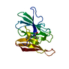
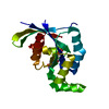
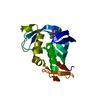
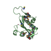
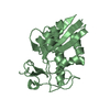
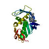
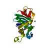
| Entry | Database: PDB / ID: 1de3 | ||||||
|---|---|---|---|---|---|---|---|
| Title | SOLUTION STRUCTURE OF THE CYTOTOXIC RIBONUCLEASE ALPHA-SARCIN | ||||||
 Components Components | RIBONUCLEASE ALPHA-SARCIN | ||||||
 Keywords Keywords | HYDROLASE / ALPHA-BETA PROTEIN | ||||||
| Function / homology |  Function and homology information Function and homology informationribotoxin / rRNA endonuclease activity / negative regulation of cytoplasmic translation / RNA endonuclease activity / hydrolase activity / RNA binding / extracellular region Similarity search - Function | ||||||
| Biological species |  | ||||||
| Method | SOLUTION NMR / torsion angle dynamics | ||||||
 Authors Authors | Perez-Canadillas, J.M. / Campos-Olivas, R. / Santoro, J. / Lacadena, J. / Martinez del Pozo, A. / Gavilanes, J.G. / Rico, M. / Bruix, M. | ||||||
 Citation Citation | Journal: J.Mol.Biol. / Year: 2000 Title: The highly refined solution structure of the cytotoxic ribonuclease alpha-sarcin reveals the structural requirements for substrate recognition and ribonucleolytic activity. Authors: Perez-Canadillas, J.M. / Santoro, J. / Campos-Olivas, R. / Lacadena, J. / Martinez del Pozo, A. / Gavilanes, J.G. / Rico, M. / Bruix, M. #1: Journal: Biochemistry / Year: 1998Title: Characterization of pKa Values and Titration Shifts in the Cytotoxic Ribonuclease alpha-Sarcin by NMR. Relationship Between Electrostatic Interactions, Structure, and Catalytic Function. Authors: Perez-Canadillas, J.M. / Campos-Olivas, R. / Lacadena, J. / Martinez del Pozo, A. / Gavilanes, J.G. / Santoro, J. / Rico, M. / Bruix, M. #2: Journal: FEBS Lett. / Year: 1996Title: Structural Basis for the Catalytic Mechanism and Substrate Specificity of the Ribonuclease alpha-Sarcin. Authors: Campos-Olivas, R. / Bruix, M. / Santoro, J. / Martinez del Pozo, A. / Lacadena, J. / Gavilanes, J.G. / Rico, M. | ||||||
| History |
|
- Structure visualization
| Structure viewer | Molecule: MolmilJmol/JSmol |
|---|
- Downloads & links
-Download
| PDBx/mmCIF format | 1de3.cif.gz | 912.9 KB | Display | PDBx/mmCIF format |
|---|---|---|---|---|
| PDB format | pdb1de3.ent.gz | 764 KB | Display | PDB format |
| PDBx/mmJSON format | 1de3.json.gz | Tree view | PDBx/mmJSON format | |
| Others |  Other downloads Other downloads |
-Validation report
| Arichive directory | https://data.pdbj.org/pub/pdb/validation_reports/de/1de3ftp://data.pdbj.org/pub/pdb/validation_reports/de/1de3 | HTTPS FTP |
|---|
-Related structure data
| Similar structure data | |
|---|---|
| Other databases |
|


-Links
 PDBj
PDBj- Assembly
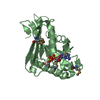
| Deposited unit | 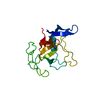
| |||||||||
|---|---|---|---|---|---|---|---|---|---|---|
| 1 |
| |||||||||
| NMR ensembles |
|
-Components
| #1: Protein | Mass: 17012.998 Da / Num. of mol.: 1 Source method: isolated from a genetically manipulated source Source: (gene. exp.)  |
|---|---|
| Has protein modification | Y |
-Experimental details
-Experiment
| Experiment | Method: SOLUTION NMR | ||||||||||||||||
|---|---|---|---|---|---|---|---|---|---|---|---|---|---|---|---|---|---|
| NMR experiment |
| ||||||||||||||||
| NMR details | Text: THIS STRUCTURE WAS DETERMINED USING STANDARD 2D HOMONUCLEAR TECHNIQUES. |
 HSQC
HSQC- Sample preparation
| Details |
| ||||||||||||
|---|---|---|---|---|---|---|---|---|---|---|---|---|---|
| Sample conditions |
| ||||||||||||
| Crystal grow | *PLUS Method: other / Details: NMR |
-NMR measurement
| NMR spectrometer | Type: Bruker AMX / Manufacturer: Bruker / Model: AMX / Field strength: 600 MHz |
|---|
- Processing
| NMR software |
| ||||||||||||||||||||||||
|---|---|---|---|---|---|---|---|---|---|---|---|---|---|---|---|---|---|---|---|---|---|---|---|---|---|
| Refinement | Method: torsion angle dynamics / Software ordinal: 1 Details: THE STRUCTURES ARE BASED ON A TOTAL OF 2777 RESTRAINTS, 2658 ARE NOE-DERIVED DISTANCE CONSTRAINTS AND 119 DIHEDRAL PHI ANGLE RESTRAINTS. THE STRUCTURES WERE ENERGY MINIMISED WITH THE GROMOS FORCE FIELD. | ||||||||||||||||||||||||
| NMR representative | Selection criteria: lowest energy | ||||||||||||||||||||||||
| NMR ensemble | Conformer selection criteria: structures with the lowest energy Conformers calculated total number: 47 / Conformers submitted total number: 20 |
