 Movie
Movie Controller

[English] 日本語
 Yorodumi
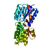
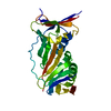
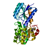
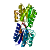
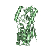
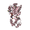
- PDB-1dbp: IDENTICAL MUTATIONS AT CORRESPONDING POSITIONS IN TWO HOMOLOGOUS ... -
+ Open data

- Basic information
| Entry | Database: PDB / ID: 1dbp | ||||||
|---|---|---|---|---|---|---|---|
| Title | IDENTICAL MUTATIONS AT CORRESPONDING POSITIONS IN TWO HOMOLOGOUS PROTEINS WITH NON-IDENTICAL EFFECTS | ||||||
 Components Components | D-RIBOSE-BINDING PROTEIN | ||||||
 Keywords Keywords | BINDING PROTEIN | ||||||
| Function / homology |  Function and homology information Function and homology informationD-ribose transmembrane transport / monosaccharide binding / positive chemotaxis / ATP-binding cassette (ABC) transporter complex, substrate-binding subunit-containing / transmembrane transport / outer membrane-bounded periplasmic space / membrane Similarity search - Function | ||||||
| Biological species |  | ||||||
| Method |  X-RAY DIFFRACTION / Resolution: 2.2 Å X-RAY DIFFRACTION / Resolution: 2.2 Å | ||||||
 Authors Authors | Mowbray, S.L. / Joakim Bjorkman, A.J. | ||||||
 Citation Citation | Journal: J.Biol.Chem. / Year: 1994 Title: Identical mutations at corresponding positions in two homologous proteins with nonidentical effects. Authors: Bjorkman, A.J. / Binnie, R.A. / Cole, L.B. / Zhang, H. / Hermodson, M.A. / Mowbray, S.L. #1: Journal: J.Mol.Biol. / Year: 1992Title: The 1.7 Angstroms X-Ray Structure of the Periplasmic Ribose Receptor from Escherichia Coli Authors: Mowbray, S.L. / Cole, L.B. #2: Journal: Protein Sci. / Year: 1992Title: Functional Mapping of the Surface of Escherichia Coli Ribose-Binding Protein: Mutations which Affect Chemotaxis and Transport Authors: Binnie, R.A. / Zhang, H. / Mowbray, S. / Hermodson, M.A. #3: Journal: J.Mol.Biol. / Year: 1990Title: Preliminary X-Ray Data for the Periplasmic Ribose Receptor from Escherichia Coli Authors: Mahendroo, M. / Cole, L.B. / Mowbray, S.L. | ||||||
| History |
|
- Structure visualization
| Structure viewer | Molecule: MolmilJmol/JSmol |
|---|
- Downloads & links
-Download
| PDBx/mmCIF format | 1dbp.cif.gz | 79.8 KB | Display | PDBx/mmCIF format |
|---|---|---|---|---|
| PDB format | pdb1dbp.ent.gz | 61.4 KB | Display | PDB format |
| PDBx/mmJSON format | 1dbp.json.gz | Tree view | PDBx/mmJSON format | |
| Others |  Other downloads Other downloads |
-Validation report
| Arichive directory | https://data.pdbj.org/pub/pdb/validation_reports/db/1dbpftp://data.pdbj.org/pub/pdb/validation_reports/db/1dbp | HTTPS FTP |
|---|
-Related structure data
| Similar structure data |
|---|


-Links
 PDBj
PDBj- Assembly
| Deposited unit | 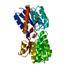
| ||||||||
|---|---|---|---|---|---|---|---|---|---|
| 1 |
| ||||||||
| Unit cell |
|
-Components
| #1: Protein | Mass: 28565.455 Da / Num. of mol.: 1 Source method: isolated from a genetically manipulated source Source: (gene. exp.) |
|---|---|
| #2: Sugar | ChemComp-RIP / 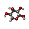  Type: D-saccharide, beta linking / Mass: 150.130 Da / Num. of mol.: 1 Type: D-saccharide, beta linking / Mass: 150.130 Da / Num. of mol.: 1Source method: isolated from a genetically manipulated source Formula: C5H10O5 |
| #3: Water | ChemComp-HOH /  Mass: 18.015 Da / Num. of mol.: 166 / Source method: isolated from a natural source / Formula: H2O Mass: 18.015 Da / Num. of mol.: 166 / Source method: isolated from a natural source / Formula: H2O |
| Sequence details | SEQUENCE ADVISORY NOTICE DIFFERENCE BETWEEN SWISS-PROT:RBSB_ECOLI AND PDB SEQUENCE. SWISS-PROT PDB ...SEQUENCE ADVISORY NOTICE DIFFERENCE |
-Experimental details
-Experiment
| Experiment | Method: X-RAY DIFFRACTION |
|---|
- Sample preparation
| Crystal | Density Matthews: 2.32 Å3/Da / Density % sol: 46.98 % | |||||||||||||||||||||||||
|---|---|---|---|---|---|---|---|---|---|---|---|---|---|---|---|---|---|---|---|---|---|---|---|---|---|---|
| Crystal grow | *PLUS pH: 7 / Method: vapor diffusion, hanging drop / Details: used to seed | |||||||||||||||||||||||||
| Components of the solutions | *PLUS
|
-Data collection
| Radiation | Scattering type: x-ray |
|---|---|
| Radiation wavelength | Relative weight: 1 |
| Reflection | *PLUS Highest resolution: 2.2 Å / Num. obs: 12590 / % possible obs: 90.2 % / Observed criterion σ(I): 11.9 / Num. measured all: 41653 / Rmerge(I) obs: 0.0698 |
- Processing
| Software |
| ||||||||||||||||||||||||||||||||||||||||||||||||||||||||||||
|---|---|---|---|---|---|---|---|---|---|---|---|---|---|---|---|---|---|---|---|---|---|---|---|---|---|---|---|---|---|---|---|---|---|---|---|---|---|---|---|---|---|---|---|---|---|---|---|---|---|---|---|---|---|---|---|---|---|---|---|---|---|
| Refinement | Resolution: 2.2→7.5 Å / σ(F): 1 /
| ||||||||||||||||||||||||||||||||||||||||||||||||||||||||||||
| Refinement step | Cycle: LAST / Resolution: 2.2→7.5 Å
| ||||||||||||||||||||||||||||||||||||||||||||||||||||||||||||
| Refine LS restraints |
| ||||||||||||||||||||||||||||||||||||||||||||||||||||||||||||
| Software | *PLUS Name: X-PLOR / Classification: refinement | ||||||||||||||||||||||||||||||||||||||||||||||||||||||||||||
| Refinement | *PLUS Rfactor obs: 0.16 / Rfactor Rwork: 0.16 | ||||||||||||||||||||||||||||||||||||||||||||||||||||||||||||
| Solvent computation | *PLUS | ||||||||||||||||||||||||||||||||||||||||||||||||||||||||||||
| Displacement parameters | *PLUS | ||||||||||||||||||||||||||||||||||||||||||||||||||||||||||||
| Refine LS restraints | *PLUS Type: x_angle_d / Dev ideal: 1.07 |
