 Movie
Movie Controller


+ Open data

- Basic information
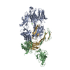
| Entry | Database: PDB / ID: 1d8s | ||||||
|---|---|---|---|---|---|---|---|
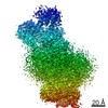
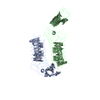
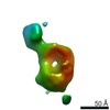
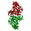
| Title | ESCHERICHIA COLI F1 ATPASE | ||||||
 Components Components |
| ||||||
 Keywords Keywords | HYDROLASE | ||||||
| Biological species |  | ||||||
| Method |  X-RAY DIFFRACTION / SYNCHROTRON / Resolution: 4.4 Å X-RAY DIFFRACTION / SYNCHROTRON / Resolution: 4.4 Å | ||||||
 Authors Authors | Hausrath, A.C. / Gruber, G. / Matthews, B.W. / Capaldi, R.A. | ||||||
 Citation Citation | Journal: Proc.Natl.Acad.Sci.USA / Year: 1999 Title: Structural features of the gamma subunit of the Escherichia coli F(1) ATPase revealed by a 4.4-A resolution map obtained by x-ray crystallography. Authors: Hausrath, A.C. / Gruber, G. / Matthews, B.W. / Capaldi, R.A. #1: Journal: FEBS Lett. / Year: 1997Title: An Improved Purification of ECF1 and ECF1F0 by Using a Cytochrome bo-Deficient Strain of Escherichia coli Facilitates Crystallization of These Complexes Authors: Gruber, G. / Hausrath, A.C. / Sagermann, M. / Capaldi, R.A. | ||||||
| History |
|
- Structure visualization
| Structure viewer | Molecule:  MolmilJmol/JSmol MolmilJmol/JSmol |
|---|
- Downloads & links
-Download
| PDBx/mmCIF format | 1d8s.cif.gz | 259.5 KB | Display | PDBx/mmCIF format |
|---|---|---|---|---|
| PDB format | pdb1d8s.ent.gz | 205.2 KB | Display | PDB format |
| PDBx/mmJSON format | 1d8s.json.gz | Tree view | PDBx/mmJSON format | |
| Others |  Other downloads Other downloads |
-Validation report
| Arichive directory | https://data.pdbj.org/pub/pdb/validation_reports/d8/1d8sftp://data.pdbj.org/pub/pdb/validation_reports/d8/1d8s | HTTPS FTP |
|---|
-Related structure data
| Similar structure data |
|---|


-Links
 PDBj
PDBj- Assembly
| Deposited unit | 
| ||||||||
|---|---|---|---|---|---|---|---|---|---|
| 1 | 
| ||||||||
| 2 | 
| ||||||||
| 3 | 
| ||||||||
| Unit cell |
|
-Components
| #1: Protein | Mass: 41889.645 Da / Num. of mol.: 3 Source method: isolated from a genetically manipulated source Source: (gene. exp.) #2: Protein | Mass: 39762.000 Da / Num. of mol.: 3 Source method: isolated from a genetically manipulated source Source: (gene. exp.) #3: Protein | | Mass: 18230.410 Da / Num. of mol.: 1 Source method: isolated from a genetically manipulated source Source: (gene. exp.) Sequence details | THIS MODEL WAS CONSTRUCTED FROM A LOW-RESOLUTION MAP. ONLY BACKBONE ATOMS HAVE BEEN MODELLED. IT IS ...THIS MODEL WAS CONSTRUCTE | |
|---|
-Experimental details
-Experiment
| Experiment | Method: X-RAY DIFFRACTION / Number of used crystals: 1 |
|---|
- Sample preparation
| Crystal | Density Matthews: 4.03 Å3/Da / Density % sol: 69.5 % | ||||||||||||||||||||||||||||||||||||||||||||||||||||||||||||||||||||||||||||||
|---|---|---|---|---|---|---|---|---|---|---|---|---|---|---|---|---|---|---|---|---|---|---|---|---|---|---|---|---|---|---|---|---|---|---|---|---|---|---|---|---|---|---|---|---|---|---|---|---|---|---|---|---|---|---|---|---|---|---|---|---|---|---|---|---|---|---|---|---|---|---|---|---|---|---|---|---|---|---|---|
| Crystal grow | Temperature: 298 K / Method: vapor diffusion, hanging drop / pH: 7.2 Details: Tris, PEG 8000, glycerol, NaCl, MgSO4, LiSO4, NaN3, EDTA, AMP-PNP, ATP, pH 7.2, VAPOR DIFFUSION, HANGING DROP, temperature 298.0K | ||||||||||||||||||||||||||||||||||||||||||||||||||||||||||||||||||||||||||||||
| Crystal grow | *PLUS Temperature: 25 ℃ | ||||||||||||||||||||||||||||||||||||||||||||||||||||||||||||||||||||||||||||||
| Components of the solutions | *PLUS
|
-Data collection
| Diffraction | Ambient pressure: 101 kPa / Mean temperature: 100 K |
|---|---|
| Diffraction source | Source: SYNCHROTRON / Site: SSRL  / Beamline: BL9-1 / Wavelength: 0.98 / Beamline: BL9-1 / Wavelength: 0.98 |
| Detector | Type: MARRESEARCH / Detector: IMAGE PLATE |
| Radiation | Protocol: SINGLE WAVELENGTH / Monochromatic (M) / Laue (L): M / Scattering type: x-ray |
| Radiation wavelength | Wavelength: 0.98 Å / Relative weight: 1 |
| Reflection | Resolution: 4.4→25 Å / Num. all: 89801 / Num. obs: 17553 / % possible obs: 64.5 % / Redundancy: 5.1 % / Rmerge(I) obs: 0.122 / Net I/σ(I): 3.7 |
| Reflection shell | Resolution: 4.4→4.44 Å / Redundancy: 5.5 % / Rmerge(I) obs: 0.358 / Num. unique all: 0 / % possible all: 53.6 |
| Reflection | *PLUS Num. measured all: 89801 |
| Reflection shell | *PLUS % possible obs: 53.6 % / Mean I/σ(I) obs: 2.1 |
- Processing
| Software |
| |||||||||||||||
|---|---|---|---|---|---|---|---|---|---|---|---|---|---|---|---|---|
| Refinement | Resolution: 4.4→25 Å / Stereochemistry target values: TNT PROTGEO 1.0 Details: Only rigid body refinement was carried out. The model consists of backbone atoms only.
| |||||||||||||||
| Refinement step | Cycle: LAST / Resolution: 4.4→25 Å
| |||||||||||||||
| Refine LS restraints |
|
