 Movie
Movie Controller

[English] 日本語
 Yorodumi
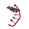
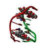
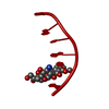
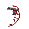
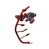
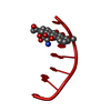
- PDB-1d58: THE MOLECULAR STRUCTURE OF A 4'-EPIADRIAMYCIN COMPLEX WITH D(TGAT... -
+ Open data

- Basic information
| Entry | Database: PDB / ID: 1d58 | ||||||||||||||||||
|---|---|---|---|---|---|---|---|---|---|---|---|---|---|---|---|---|---|---|---|
| Title | THE MOLECULAR STRUCTURE OF A 4'-EPIADRIAMYCIN COMPLEX WITH D(TGATCA) AT 1.7 ANGSTROM RESOLUTION-COMPARISON WITH THE STRUCTURE OF 4'-EPIADRIAMYCIN D(TGTACA) AND D(CGATCG) COMPLEXES | ||||||||||||||||||
 Components Components | DNA (5'-D(* Keywords KeywordsDNA / RIGHT HANDED DNA / DOUBLE HELIX / COMPLEXED WITH DRUG | Function / homology | 4'-EPIDOXORUBICIN / DNA |  Function and homology information Function and homology informationMethod |  X-RAY DIFFRACTION / Resolution: 1.7 Å X-RAY DIFFRACTION / Resolution: 1.7 Å  Authors AuthorsLanglois D'Estaintot, B. / Gallois, B. / Brown, T. / Hunter, W.N. |  CitationJournal: Nucleic Acids Res. / Year: 1992 CitationJournal: Nucleic Acids Res. / Year: 1992Title: The molecular structure of a 4'-epiadriamycin complex with d(TGATCA) at 1.7A resolution: comparison with the structure of 4'-epiadriamycin d(TGTACA) and d(CGATCG) complexes. Authors: Langlois d'Estaintot, B. / Gallois, B. / Brown, T. / Hunter, W.N. History |
|
- Structure visualization
| Structure viewer | Molecule: MolmilJmol/JSmol |
|---|
- Downloads & links
-Download
| PDBx/mmCIF format | 1d58.cif.gz | 14.1 KB | Display | PDBx/mmCIF format |
|---|---|---|---|---|
| PDB format | pdb1d58.ent.gz | 7.9 KB | Display | PDB format |
| PDBx/mmJSON format | 1d58.json.gz | Tree view | PDBx/mmJSON format | |
| Others |  Other downloads Other downloads |
-Validation report
| Summary document | 1d58_validation.pdf.gz | 737.4 KB | Display | wwPDB validaton report |
|---|---|---|---|---|
| Full document | 1d58_full_validation.pdf.gz | 743.3 KB | Display | |
| Data in XML | 1d58_validation.xml.gz | 4.2 KB | Display | |
| Data in CIF | 1d58_validation.cif.gz | 4.7 KB | Display | |
| Arichive directory | https://data.pdbj.org/pub/pdb/validation_reports/d5/1d58ftp://data.pdbj.org/pub/pdb/validation_reports/d5/1d58 | HTTPS FTP |
-Related structure data
| Similar structure data |
|---|


-Links
 PDBj
PDBj

- Assembly
| Deposited unit | 
| ||||||||
|---|---|---|---|---|---|---|---|---|---|
| 1 | 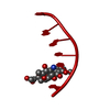
| ||||||||
| Unit cell |
|
-Components
| #1: DNA chain | Mass: 1808.229 Da / Num. of mol.: 1 / Source method: obtained synthetically |
|---|---|
| #2: Chemical | ChemComp-DM6 / 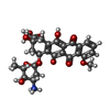  Mass: 544.527 Da / Num. of mol.: 1 / Source method: obtained synthetically / Formula: C27H30NO11 / Comment: medication, chemotherapy*YM Mass: 544.527 Da / Num. of mol.: 1 / Source method: obtained synthetically / Formula: C27H30NO11 / Comment: medication, chemotherapy*YM |
| #3: Water | ChemComp-HOH /  Mass: 18.015 Da / Num. of mol.: 34 / Source method: isolated from a natural source / Formula: H2O Mass: 18.015 Da / Num. of mol.: 34 / Source method: isolated from a natural source / Formula: H2O |
-Experimental details
-Experiment
| Experiment | Method: X-RAY DIFFRACTION |
|---|
- Sample preparation
| Crystal | Density Matthews: 2.87 Å3/Da / Density % sol: 57.18 % | ||||||||||||||||||||||||||||||||||||||||
|---|---|---|---|---|---|---|---|---|---|---|---|---|---|---|---|---|---|---|---|---|---|---|---|---|---|---|---|---|---|---|---|---|---|---|---|---|---|---|---|---|---|
| Crystal grow | Temperature: 277 K / Method: vapor diffusion / pH: 6.5 / Details: pH 6.50, VAPOR DIFFUSION, temperature 277.00K | ||||||||||||||||||||||||||||||||||||||||
| Components of the solutions |
| ||||||||||||||||||||||||||||||||||||||||
| Crystal grow | *PLUS Temperature: 4 ℃ / Method: vapor diffusion, sitting drop / pH: 6.5 | ||||||||||||||||||||||||||||||||||||||||
| Components of the solutions | *PLUS
|
-Data collection
| Diffraction | Mean temperature: 295 K |
|---|---|
| Diffraction source | Source: ROTATING ANODE / Type: RIGAKU RU200 |
| Detector | Type: RIGAKU AFC-5 / Detector: DIFFRACTOMETER |
| Radiation | Scattering type: x-ray |
| Radiation wavelength | Relative weight: 1 |
| Reflection | Highest resolution: 1.7 Å / Num. all: 10067 / Num. obs: 5730 |
| Reflection | *PLUS Highest resolution: 1.7 Å / Rmerge(I) obs: 0.102 |
- Processing
| Software | Name: X-PLOR / Classification: refinement | ||||||||||||||||
|---|---|---|---|---|---|---|---|---|---|---|---|---|---|---|---|---|---|
| Refinement | Resolution: 1.7→7 Å / σ(F): 2 /
| ||||||||||||||||
| Refine Biso |
| ||||||||||||||||
| Refinement step | Cycle: LAST / Resolution: 1.7→7 Å
| ||||||||||||||||
| Software | *PLUS Name: X-PLOR / Classification: refinement | ||||||||||||||||
| Refinement | *PLUS Highest resolution: 1.7 Å / Lowest resolution: 7 Å / σ(F): 2 / Rfactor obs: 0.202 | ||||||||||||||||
| Solvent computation | *PLUS | ||||||||||||||||
| Displacement parameters | *PLUS |
