 Movie
Movie Controller

[English] 日本語
 Yorodumi
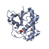
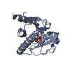
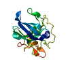
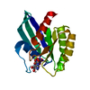
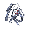
- PDB-1d1p: CRYSTAL STRUCTURE OF A YEAST LOW MOLECULAR WEIGHT PROTEIN TYROSIN... -
+ Open data

- Basic information
| Entry | Database: PDB / ID: 1d1p | ||||||
|---|---|---|---|---|---|---|---|
| Title | CRYSTAL STRUCTURE OF A YEAST LOW MOLECULAR WEIGHT PROTEIN TYROSINE PHOSPHATASE (LTP1) | ||||||
 Components Components | TYROSINE PHOSPHATASE | ||||||
 Keywords Keywords | HYDROLASE / BETA-ALPHA-BETA / TYROSINE PHOSPHATASE / LTP1 | ||||||
| Function / homology |  Function and homology information Function and homology informationacid phosphatase / acid phosphatase activity / protein-tyrosine-phosphatase / protein tyrosine phosphatase activity / nucleus / cytoplasm Similarity search - Function | ||||||
| Biological species |  | ||||||
| Method |  X-RAY DIFFRACTION / MOLECULAR REPLACEMENT / Resolution: 2.2 Å X-RAY DIFFRACTION / MOLECULAR REPLACEMENT / Resolution: 2.2 Å | ||||||
 Authors Authors | Wang, S. / Tabernero, L. / Zhang, M. / Harms, E. / Van Etten, R.L. / Stauffacher, C.V. | ||||||
 Citation Citation | Journal: Biochemistry / Year: 2000 Title: Crystal structures of a low-molecular weight protein tyrosine phosphatase from Saccharomyces cerevisiae and its complex with the substrate p-nitrophenyl phosphate. Authors: Wang, S. / Tabernero, L. / Zhang, M. / Harms, E. / Van Etten, R.L. / Stauffacher, C.V. #1: Journal: J.Biol.Chem. / Year: 1995Title: Cloning and Characterization of a Saccharomyces cerevisiae Gene Encoding the Low Molecular Weight Protein-tyrosine Phosphatase Authors: Ostanin, K. / Pokalsky, C. / Wang, S. / Van Etten, R.L. | ||||||
| History |
|
- Structure visualization
| Structure viewer | Molecule: MolmilJmol/JSmol |
|---|
- Downloads & links
-Download
| PDBx/mmCIF format | 1d1p.cif.gz | 78.5 KB | Display | PDBx/mmCIF format |
|---|---|---|---|---|
| PDB format | pdb1d1p.ent.gz | 58.5 KB | Display | PDB format |
| PDBx/mmJSON format | 1d1p.json.gz | Tree view | PDBx/mmJSON format | |
| Others |  Other downloads Other downloads |
-Validation report
| Arichive directory | https://data.pdbj.org/pub/pdb/validation_reports/d1/1d1pftp://data.pdbj.org/pub/pdb/validation_reports/d1/1d1p | HTTPS FTP |
|---|
-Related structure data
| Related structure data | 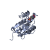 1d1qC 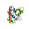 1pntS C: citing same article ( S: Starting model for refinement |
|---|---|
| Similar structure data |


-Links
 PDBj
PDBj

- Assembly
| Deposited unit | 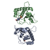
| ||||||||
|---|---|---|---|---|---|---|---|---|---|
| 1 | 
| ||||||||
| 2 | 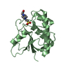
| ||||||||
| Unit cell |
| ||||||||
| Noncrystallographic symmetry (NCS) | NCS oper: (Code: given Matrix: (-0.156013, -0.737096, -0.657533), Vector: |
-Components
| #1: Protein | Mass: 18572.025 Da / Num. of mol.: 2 Source method: isolated from a genetically manipulated source Source: (gene. exp.) Plasmid: PT7-7 / Production host:  #2: Chemical |   Mass: 238.305 Da / Num. of mol.: 2 / Source method: obtained synthetically / Formula: C8H18N2O4S / Comment: pH buffer*YM Mass: 238.305 Da / Num. of mol.: 2 / Source method: obtained synthetically / Formula: C8H18N2O4S / Comment: pH buffer*YM#3: Water | ChemComp-HOH / |  Mass: 18.015 Da / Num. of mol.: 106 / Source method: isolated from a natural source / Formula: H2O Mass: 18.015 Da / Num. of mol.: 106 / Source method: isolated from a natural source / Formula: H2O |
|---|
-Experimental details
-Experiment
| Experiment | Method: X-RAY DIFFRACTION / Number of used crystals: 2 |
|---|
- Sample preparation
| Crystal | Density Matthews: 2.19 Å3/Da / Density % sol: 43.8 % | ||||||||||||||||||||||||||||||
|---|---|---|---|---|---|---|---|---|---|---|---|---|---|---|---|---|---|---|---|---|---|---|---|---|---|---|---|---|---|---|---|
| Crystal grow | Temperature: 298 K / Method: vapor diffusion, sitting drop / pH: 7 Details: PEG 3400, HEPES, sodium chloride, pH 7.0, VAPOR DIFFUSION, SITTING DROP, temperature 298.0K | ||||||||||||||||||||||||||||||
| Crystal grow | *PLUS | ||||||||||||||||||||||||||||||
| Components of the solutions | *PLUS
|
-Data collection
| Diffraction | Mean temperature: 298 K |
|---|---|
| Diffraction source | Source: ROTATING ANODE / Type: RIGAKU RU200 / Wavelength: 1.5418 |
| Detector | Type: RIGAKU RAXIS II / Detector: IMAGE PLATE / Date: Nov 12, 1995 / Details: double focussing mirror |
| Radiation | Protocol: SINGLE WAVELENGTH / Monochromatic (M) / Laue (L): M / Scattering type: x-ray |
| Radiation wavelength | Wavelength: 1.5418 Å / Relative weight: 1 |
| Reflection | Resolution: 2.2→30 Å / Num. all: 15614 / Num. obs: 15614 / % possible obs: 91.1 % / Observed criterion σ(I): 0 / Redundancy: 9 % / Biso Wilson estimate: 11.9 Å2 / Rmerge(I) obs: 0.058 / Rsym value: 0.058 / Net I/σ(I): 25.9 |
| Reflection shell | Resolution: 2.2→2.28 Å / Redundancy: 2.5 % / Rmerge(I) obs: 0.204 / Mean I/σ(I) obs: 7.28 / Num. unique all: 1414 / Rsym value: 0.204 / % possible all: 84.6 |
| Reflection | *PLUS Num. measured all: 140622 |
| Reflection shell | *PLUS % possible obs: 84.6 % |
- Processing
| Software |
| ||||||||||||||||||||||||||||||||||||||||||||||||||||||||||||
|---|---|---|---|---|---|---|---|---|---|---|---|---|---|---|---|---|---|---|---|---|---|---|---|---|---|---|---|---|---|---|---|---|---|---|---|---|---|---|---|---|---|---|---|---|---|---|---|---|---|---|---|---|---|---|---|---|---|---|---|---|---|
| Refinement | Method to determine structure: MOLECULAR REPLACEMENT Starting model: Bovine low molecular weight protein tyrosine phosphatase (BPTP) PDB ID: 1PNT Resolution: 2.2→12 Å / Rfactor Rfree error: 0.007 / Data cutoff high absF: 10000000 / Data cutoff low absF: 0 / Isotropic thermal model: RESTRAINED / Cross valid method: THROUGHOUT / σ(F): 0 / Stereochemistry target values: Engh & Huber / Details: bulk solvent model used
| ||||||||||||||||||||||||||||||||||||||||||||||||||||||||||||
| Displacement parameters | Biso mean: 25.9 Å2
| ||||||||||||||||||||||||||||||||||||||||||||||||||||||||||||
| Refine analyze |
| ||||||||||||||||||||||||||||||||||||||||||||||||||||||||||||
| Refinement step | Cycle: LAST / Resolution: 2.2→12 Å
| ||||||||||||||||||||||||||||||||||||||||||||||||||||||||||||
| Refine LS restraints |
| ||||||||||||||||||||||||||||||||||||||||||||||||||||||||||||
| Refine LS restraints NCS | NCS model details: RESTRAINED | ||||||||||||||||||||||||||||||||||||||||||||||||||||||||||||
| LS refinement shell | Resolution: 2.2→2.34 Å / Rfactor Rfree error: 0.026 / Total num. of bins used: 6
| ||||||||||||||||||||||||||||||||||||||||||||||||||||||||||||
| Xplor file |
| ||||||||||||||||||||||||||||||||||||||||||||||||||||||||||||
| Software | *PLUS Name: X-PLOR / Version: 3.851 / Classification: refinement | ||||||||||||||||||||||||||||||||||||||||||||||||||||||||||||
| Refine LS restraints | *PLUS
|
