 Movie
Movie Controller

[English] 日本語
 Yorodumi
- PDB-1cx1: SECOND N-TERMINAL CELLULOSE-BINDING DOMAIN FROM CELLULOMONAS FIMI... -
+ Open data

- Basic information
| Entry | Database: PDB / ID: 1cx1 | ||||||
|---|---|---|---|---|---|---|---|
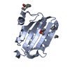
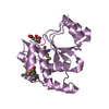
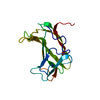
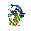
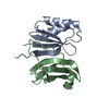
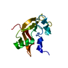
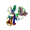
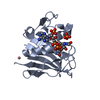
| Title | SECOND N-TERMINAL CELLULOSE-BINDING DOMAIN FROM CELLULOMONAS FIMI BETA-1,4-GLUCANASE C, NMR, 22 STRUCTURES | ||||||
 Components Components | ENDOGLUCANASE C | ||||||
 Keywords Keywords | HYDROLASE / CELLULOSE-BINDING DOMAIN / CELLOOLIGOSACHARIDES / CELLULASE / PROTEIN- CARBOHYDRATE INTERACTION | ||||||
| Function / homology |  Function and homology information Function and homology information | ||||||
| Biological species |  Cellulomonas fimi (bacteria) Cellulomonas fimi (bacteria) | ||||||
| Method | SOLUTION NMR / ALL CALCULATIONS WERE PERFORMED USING X-PLOR V3.8 WITH SCRIPTS FROM THE X-PLOR 3.1 MANUAL A. A. BRUNGER (1992), NEW HAVEN: YALE UNIVERSITY PRESS ACCORDING TO THE METHOD DESCRIBED BY M. NILGES, G. M. CLORE, A. M. GRONENBORN | ||||||
| Model type details | minimized average | ||||||
 Authors Authors | Brun, E. / Johnson, P.E. / Creagh, L.A. / Haynes, C.A. / Tomme, P. / Webster, P. / Kilburn, D.G. / McIntosh, L.P. | ||||||
 Citation Citation | Journal: Biochemistry / Year: 2000 Title: Structure and binding specificity of the second N-terminal cellulose-binding domain from Cellulomonas fimi endoglucanase C. Authors: Brun, E. / Johnson, P.E. / Creagh, A.L. / Tomme, P. / Webster, P. / Haynes, C.A. / McIntosh, L.P. #1: Journal: J.Mol.Biol. / Year: 1999Title: The cellulose-binding domains from Cellulomonas fimi beta-1, 4-glucanase CenC bind nitroxide spin-labeled cellooligosaccharides in multiple orientations Authors: Brun, E. / Johnson, P.E. / MacKenzie, L. / Withers, S.G. / McIntosh, L.P. #2: Journal: Biochemistry / Year: 1997Title: Structure of the N-terminal Cellulose-Binding Domain of Cellulomonas Fimi CenC Determined by Nucelar Magnetic Resonance Spectroscopy Authors: Johnson, P.E. / Joshi, M.D. / Tomme, P. / Kilburn, D.G. / McIntosh, L.P. | ||||||
| History |
|
- Structure visualization
| Structure viewer | Molecule: MolmilJmol/JSmol |
|---|
- Downloads & links
-Download
| PDBx/mmCIF format | 1cx1.cif.gz | 1 MB | Display | PDBx/mmCIF format |
|---|---|---|---|---|
| PDB format | pdb1cx1.ent.gz | 875.3 KB | Display | PDB format |
| PDBx/mmJSON format | 1cx1.json.gz | Tree view | PDBx/mmJSON format | |
| Others |  Other downloads Other downloads |
-Validation report
| Arichive directory | https://data.pdbj.org/pub/pdb/validation_reports/cx/1cx1ftp://data.pdbj.org/pub/pdb/validation_reports/cx/1cx1 | HTTPS FTP |
|---|
-Related structure data
| Related structure data | |
|---|---|
| Similar structure data |


-Links
 PDBj
PDBj


- Assembly
| Deposited unit | 
| |||||||||
|---|---|---|---|---|---|---|---|---|---|---|
| 1 |
| |||||||||
| NMR ensembles |
|
-Components
| #1: Protein | Mass: 15877.379 Da / Num. of mol.: 1 / Fragment: RESIDUES 176-328 Source method: isolated from a genetically manipulated source Source: (gene. exp.) Cellulomonas fimi (bacteria) / Production host: |
|---|---|
| Has protein modification | Y |
-Experimental details
-Experiment
| Experiment | Method: SOLUTION NMR | ||||||||||||||||||||||||
|---|---|---|---|---|---|---|---|---|---|---|---|---|---|---|---|---|---|---|---|---|---|---|---|---|---|
| NMR experiment |
| ||||||||||||||||||||||||
| NMR details | Text: CBDN2 WAS STUDIED IN THE PRESENCE OF SATURATING CONCENTRATIONS OF CELLOPENTAOSE. HOWEVER, DUE TO SPECTRAL OVERLAP, THE OLIGOSACCHARIDE WAS NOT INCLUDED IN THE STRUCTURE CALCULATIONS |
- Sample preparation
| Details |
| ||||||||
|---|---|---|---|---|---|---|---|---|---|
| Sample conditions | Ionic strength: 50mMNACL / pH: 6 / Pressure: 1 atm / Temperature: 35 K | ||||||||
| Crystal grow | *PLUS Method: other / Details: NMR |
-NMR measurement
| NMR spectrometer | Type: Varian UNITY / Manufacturer: Varian / Model: UNITY / Field strength: 500 MHz |
|---|
- Processing
| NMR software |
| ||||||||||||||||||||||||
|---|---|---|---|---|---|---|---|---|---|---|---|---|---|---|---|---|---|---|---|---|---|---|---|---|---|
| Refinement | Method: ALL CALCULATIONS WERE PERFORMED USING X-PLOR V3.8 WITH SCRIPTS FROM THE X-PLOR 3.1 MANUAL A. A. BRUNGER (1992), NEW HAVEN: YALE UNIVERSITY PRESS ACCORDING TO THE METHOD DESCRIBED BY M. ...Method: ALL CALCULATIONS WERE PERFORMED USING X-PLOR V3.8 WITH SCRIPTS FROM THE X-PLOR 3.1 MANUAL A. A. BRUNGER (1992), NEW HAVEN: YALE UNIVERSITY PRESS ACCORDING TO THE METHOD DESCRIBED BY M. NILGES, G. M. CLORE, A. M. GRONENBORN Software ordinal: 1 Details: A TOTAL OF 1510 NOE DERIVED DISTANCE RESTRAINTS THAT COMPRISE 700 INTRARESIDUE, 332 SEQUENTIAL, 83 SHORT RANGE (1<|I-J|<=4), 394 LONG RANGE (|I-J|>4) WERE INCLUDED.. 1 DISULPHIDE BOND DISTANCE RESTRAINT, 86 HYDROGEN BOND RESTRAINTS, 53 PHI ANGLE RESTRAINTS, 70 PSI ANGLE RESTRAINTS, AND 48 CHI1 RESTRAINTS WERE ALSO INCLUDED. | ||||||||||||||||||||||||
| NMR representative | Selection criteria: minimized average structure | ||||||||||||||||||||||||
| NMR ensemble | Conformer selection criteria: STRUCTURES WITH ACCEPTABLE COVALENT GEOMETRY,STRUCTURES WITH THE LEAST RESTRAINT VIOLATIONS,STRUCTURES WITH THE LOWEST ENERGY Conformers calculated total number: 60 / Conformers submitted total number: 22 |
 X-PLOR