 Movie
Movie Controller

[English] 日本語
 Yorodumi
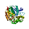
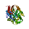
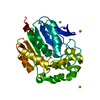
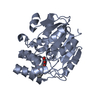
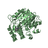
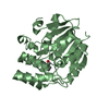
- PDB-1cv2: Hydrolytic haloalkane dehalogenase linb from sphingomonas paucimo... -
+ Open data

- Basic information
| Entry | Database: PDB / ID: 1cv2 | ||||||
|---|---|---|---|---|---|---|---|
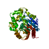
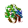
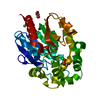
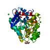
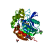
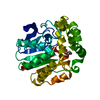
| Title | Hydrolytic haloalkane dehalogenase linb from sphingomonas paucimobilis UT26 AT 1.6 A resolution | ||||||
 Components Components | HALOALKANE DEHALOGENASE | ||||||
 Keywords Keywords | HYDROLASE / DEHALOGENASE / LINDANE / BIODEGRADATION / ALPHA/BETA-HYDROLASE | ||||||
| Function / homology |  Function and homology information Function and homology informationhaloalkane dehalogenase / haloalkane dehalogenase activity / response to toxic substance / periplasmic space Similarity search - Function | ||||||
| Biological species |  Sphingomonas paucimobilis (bacteria) Sphingomonas paucimobilis (bacteria) | ||||||
| Method |  X-RAY DIFFRACTION / SYNCHROTRON / MOLECULAR REPLACEMENT / Resolution: 1.58 Å X-RAY DIFFRACTION / SYNCHROTRON / MOLECULAR REPLACEMENT / Resolution: 1.58 Å | ||||||
 Authors Authors | Marek, J. / Vevodova, J. / Damborsky, J. / Smatanova, I. / Svensson, L.A. / Newman, J. / Nagata, Y. / Takagi, M. | ||||||
 Citation Citation | Journal: Biochemistry / Year: 2000 Title: Crystal structure of the haloalkane dehalogenase from Sphingomonas paucimobilis UT26. Authors: Marek, J. / Vevodova, J. / Smatanova, I.K. / Nagata, Y. / Svensson, L.A. / Newman, J. / Takagi, M. / Damborsky, J. #1: Journal: Acta Crystallogr.,Sect.D / Year: 1999Title: Crystallization and Preliminary X-Ray Diffraction Analysis of Haloalkane Dehalogenase Linb from Sphingomonas Paucimobilis Ut26 Authors: Smatanova, I. / Nagata, Y. / Svensson, L.A. / Takagi, M. / Marek, J. | ||||||
| History |
|
- Structure visualization
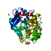
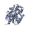
| Structure viewer | Molecule: MolmilJmol/JSmol |
|---|
- Downloads & links
-Download
| PDBx/mmCIF format | 1cv2.cif.gz | 80.8 KB | Display | PDBx/mmCIF format |
|---|---|---|---|---|
| PDB format | pdb1cv2.ent.gz | 58.2 KB | Display | PDB format |
| PDBx/mmJSON format | 1cv2.json.gz | Tree view | PDBx/mmJSON format | |
| Others |  Other downloads Other downloads |
-Validation report
| Summary document | 1cv2_validation.pdf.gz | 425.6 KB | Display | wwPDB validaton report |
|---|---|---|---|---|
| Full document | 1cv2_full_validation.pdf.gz | 431.7 KB | Display | |
| Data in XML | 1cv2_validation.xml.gz | 17.9 KB | Display | |
| Data in CIF | 1cv2_validation.cif.gz | 27.9 KB | Display | |
| Arichive directory | https://data.pdbj.org/pub/pdb/validation_reports/cv/1cv2ftp://data.pdbj.org/pub/pdb/validation_reports/cv/1cv2 | HTTPS FTP |
-Related structure data
| Related structure data | 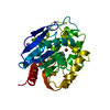 1d07C 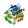 1bn6S S: Starting model for refinement C: citing same article ( |
|---|---|
| Similar structure data |


-Links
 PDBj
PDBj


- Assembly
| Deposited unit | 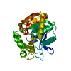
| ||||||||||
|---|---|---|---|---|---|---|---|---|---|---|---|
| 1 |
| ||||||||||
| Unit cell |
| ||||||||||
| Components on special symmetry positions |
|
-Components
| #1: Protein | Mass: 33144.609 Da / Num. of mol.: 1 Source method: isolated from a genetically manipulated source Source: (gene. exp.) Sphingomonas paucimobilis (bacteria) / Strain: UT26 / Plasmid details: TAC PROMOTER SYSTEM / Plasmid: PMYLB1 / Production host: References: UniProt: P51698, UniProt: D4Z2G1*PLUS, haloalkane dehalogenase |
|---|---|
| #2: Water | ChemComp-HOH /  Mass: 18.015 Da / Num. of mol.: 449 / Source method: isolated from a natural source / Formula: H2O Mass: 18.015 Da / Num. of mol.: 449 / Source method: isolated from a natural source / Formula: H2O |
-Experimental details
-Experiment
| Experiment | Method: X-RAY DIFFRACTION / Number of used crystals: 1 |
|---|
- Sample preparation
| Crystal | Density Matthews: 2.07 Å3/Da / Density % sol: 40.2 % | ||||||||||||||||||||
|---|---|---|---|---|---|---|---|---|---|---|---|---|---|---|---|---|---|---|---|---|---|
| Crystal grow | Temperature: 278 K / Method: vapor diffusion, hanging drop / pH: 8.9 Details: 19% (W/V) PEG 6000, 100 MM TRIS-HCL, 200MM CA ACETATE, PH=8.9, VAPOR DIFFUSION, HANGING DROP at 278K | ||||||||||||||||||||
| Crystal grow | *PLUS Details: microseeding | ||||||||||||||||||||
| Components of the solutions | *PLUS
|
-Data collection
| Diffraction | Mean temperature: 100 K |
|---|---|
| Diffraction source | Source: SYNCHROTRON / Site: MAX II  / Beamline: I711 / Wavelength: 0.942 / Beamline: I711 / Wavelength: 0.942 |
| Detector | Type: MARRESEARCH / Detector: IMAGE PLATE / Date: Nov 18, 1998 |
| Radiation | Protocol: SINGLE WAVELENGTH / Monochromatic (M) / Laue (L): M / Scattering type: x-ray |
| Radiation wavelength | Wavelength: 0.942 Å / Relative weight: 1 |
| Reflection | Resolution: 1.575→23.75 Å / Num. all: 34649 / Num. obs: 34649 / % possible obs: 94.2 % / Redundancy: 4.84 % / Biso Wilson estimate: 11.5 Å2 / Rmerge(I) obs: 0.065 / Net I/σ(I): 18.31 |
| Reflection shell | Resolution: 1.575→1.6 Å / Redundancy: 2.79 % / Rmerge(I) obs: 0.146 / Mean I/σ(I) obs: 5.98 / % possible all: 81.4 |
| Reflection | *PLUS Highest resolution: 1.58 Å / Lowest resolution: 20 Å / Num. obs: 34513 |
| Reflection shell | *PLUS % possible obs: 81.4 % |
- Processing
| Software |
| |||||||||||||||||||||||||||||||||
|---|---|---|---|---|---|---|---|---|---|---|---|---|---|---|---|---|---|---|---|---|---|---|---|---|---|---|---|---|---|---|---|---|---|---|
| Refinement | Method to determine structure: MOLECULAR REPLACEMENT Starting model: 1BN6 Resolution: 1.58→20 Å / Rfactor Rfree error: 0.005 / Num. parameters: 1100 / Num. restraintsaints: 958 / Cross valid method: THROUGHOUT / σ(F): 0
| |||||||||||||||||||||||||||||||||
| Solvent computation | Solvent model: MOEWS & KRETSINGER, J.MOL.BIOL.91(1973)201-228 | |||||||||||||||||||||||||||||||||
| Displacement parameters | Biso mean: 14.7 Å2 | |||||||||||||||||||||||||||||||||
| Refine analyze | Occupancy sum hydrogen: 0 / Occupancy sum non hydrogen: 2749.5
| |||||||||||||||||||||||||||||||||
| Refinement step | Cycle: LAST / Resolution: 1.58→20 Å
| |||||||||||||||||||||||||||||||||
| Refine LS restraints |
| |||||||||||||||||||||||||||||||||
| LS refinement shell | Resolution: 1.58→1.68 Å / Rfactor Rfree error: 0.014
| |||||||||||||||||||||||||||||||||
| Software | *PLUS Name: SHELXL-97 / Classification: refinement | |||||||||||||||||||||||||||||||||
| Refinement | *PLUS Rfactor Rfree: 0.204 / Rfactor Rwork: 0.145 | |||||||||||||||||||||||||||||||||
| Solvent computation | *PLUS | |||||||||||||||||||||||||||||||||
| Displacement parameters | *PLUS | |||||||||||||||||||||||||||||||||
| Refine LS restraints | *PLUS
|
