 Movie
Movie Controller

[English] 日本語
 Yorodumi
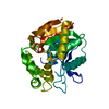
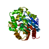
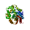
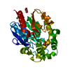
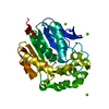
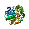
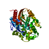
- PDB-4h7i: Crystal structure of haloalkane dehalogenase LinB L138I mutant fr... -
+ Open data

- Basic information
| Entry | Database: PDB / ID: 4h7i | ||||||
|---|---|---|---|---|---|---|---|
| Title | Crystal structure of haloalkane dehalogenase LinB L138I mutant from Sphingobium sp. MI1205 | ||||||
 Components Components | Haloalkane dehalogenase | ||||||
 Keywords Keywords | HYDROLASE / alpha/beta hydrolase fold | ||||||
| Function / homology |  Function and homology information Function and homology informationhaloalkane dehalogenase / haloalkane dehalogenase activity / metal ion binding Similarity search - Function | ||||||
| Biological species |  Sphingobium (bacteria) Sphingobium (bacteria) | ||||||
| Method |  X-RAY DIFFRACTION / SYNCHROTRON / MOLECULAR REPLACEMENT / Resolution: 1.8 Å X-RAY DIFFRACTION / SYNCHROTRON / MOLECULAR REPLACEMENT / Resolution: 1.8 Å | ||||||
 Authors Authors | Okai, M. / Ohtsuka, J. / Imai, L.F. / Mase, T. / Moriuchi, R. / Tsuda, M. / Nagata, K. / Nagata, Y. / Tanokura, M. | ||||||
 Citation Citation | Journal: J.Bacteriol. / Year: 2013 Title: Crystal Structure and Site-Directed Mutagenesis Analyses of Haloalkane Dehalogenase LinB from Sphingobium sp. Strain MI1205. Authors: Okai, M. / Ohtsuka, J. / Imai, L.F. / Mase, T. / Moriuchi, R. / Tsuda, M. / Nagata, K. / Nagata, Y. / Tanokura, M. | ||||||
| History |
|
- Structure visualization
| Structure viewer | Molecule: MolmilJmol/JSmol |
|---|
- Downloads & links
-Download
| PDBx/mmCIF format | 4h7i.cif.gz | 75.4 KB | Display | PDBx/mmCIF format |
|---|---|---|---|---|
| PDB format | pdb4h7i.ent.gz | 54.7 KB | Display | PDB format |
| PDBx/mmJSON format | 4h7i.json.gz | Tree view | PDBx/mmJSON format | |
| Others |  Other downloads Other downloads |
-Validation report
| Arichive directory | https://data.pdbj.org/pub/pdb/validation_reports/h7/4h7iftp://data.pdbj.org/pub/pdb/validation_reports/h7/4h7i | HTTPS FTP |
|---|
-Related structure data
| Related structure data | 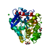 4h77SC 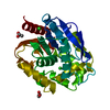 4h7dC 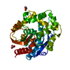 4h7eC 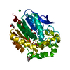 4h7fC 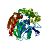 4h7hC  4h7jC 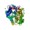 4h7kC S: Starting model for refinement C: citing same article ( |
|---|---|
| Similar structure data |


-Links
 PDBj
PDBj


- Assembly
| Deposited unit | 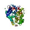
| ||||||||
|---|---|---|---|---|---|---|---|---|---|
| 1 |
| ||||||||
| Unit cell |
| ||||||||
| Components on special symmetry positions |
|
-Components
| #1: Protein | Mass: 34096.586 Da / Num. of mol.: 1 / Mutation: L138I Source method: isolated from a genetically manipulated source Source: (gene. exp.) Sphingobium (bacteria) / Strain: MI1205 / Gene: linB, dhaA / Production host: | ||||
|---|---|---|---|---|---|
| #2: Chemical | ChemComp-CA /   Mass: 40.078 Da / Num. of mol.: 1 / Source method: obtained synthetically / Formula: Ca Mass: 40.078 Da / Num. of mol.: 1 / Source method: obtained synthetically / Formula: Ca | ||||
| #3: Chemical |   Mass: 35.453 Da / Num. of mol.: 2 / Source method: obtained synthetically / Formula: Cl Mass: 35.453 Da / Num. of mol.: 2 / Source method: obtained synthetically / Formula: Cl#4: Chemical |   Mass: 92.094 Da / Num. of mol.: 2 / Source method: obtained synthetically / Formula: C3H8O3 Mass: 92.094 Da / Num. of mol.: 2 / Source method: obtained synthetically / Formula: C3H8O3#5: Water | ChemComp-HOH / |  Mass: 18.015 Da / Num. of mol.: 206 / Source method: isolated from a natural source / Formula: H2O Mass: 18.015 Da / Num. of mol.: 206 / Source method: isolated from a natural source / Formula: H2O |
-Experimental details
-Experiment
| Experiment | Method: X-RAY DIFFRACTION / Number of used crystals: 1 |
|---|
- Sample preparation
| Crystal | Density Matthews: 1.98 Å3/Da / Density % sol: 37.74 % |
|---|---|
| Crystal grow | Temperature: 278 K / Method: vapor diffusion, sitting drop / Details: VAPOR DIFFUSION, SITTING DROP, temperature 278K |
-Data collection
| Diffraction source | Source: SYNCHROTRON / Site: Photon Factory  / Beamline: BL-5A / Wavelength: 1 Å / Beamline: BL-5A / Wavelength: 1 Å |
|---|---|
| Detector | Date: Jun 25, 2010 |
| Radiation | Protocol: SINGLE WAVELENGTH / Monochromatic (M) / Laue (L): M / Scattering type: x-ray |
| Radiation wavelength | Wavelength: 1 Å / Relative weight: 1 |
| Reflection | Resolution: 1.8→20 Å / Num. obs: 25640 |
- Processing
| Software |
| |||||||||||||||||||||||||||||||||||||||||||||
|---|---|---|---|---|---|---|---|---|---|---|---|---|---|---|---|---|---|---|---|---|---|---|---|---|---|---|---|---|---|---|---|---|---|---|---|---|---|---|---|---|---|---|---|---|---|---|
| Refinement | Method to determine structure: MOLECULAR REPLACEMENT Starting model: 4H77 Resolution: 1.8→20 Å / Cor.coef. Fo:Fc: 0.954 / Cor.coef. Fo:Fc free: 0.941 / SU B: 2.161 / SU ML: 0.069 / Cross valid method: THROUGHOUT / ESU R: 0.134 / ESU R Free: 0.114 / Stereochemistry target values: MAXIMUM LIKELIHOOD / Details: HYDROGENS HAVE BEEN USED IF PRESENT IN THE INPUT
| |||||||||||||||||||||||||||||||||||||||||||||
| Solvent computation | Ion probe radii: 0.8 Å / Shrinkage radii: 0.8 Å / VDW probe radii: 1.2 Å / Solvent model: MASK | |||||||||||||||||||||||||||||||||||||||||||||
| Displacement parameters | Biso mean: 13.66 Å2
| |||||||||||||||||||||||||||||||||||||||||||||
| Refinement step | Cycle: LAST / Resolution: 1.8→20 Å
| |||||||||||||||||||||||||||||||||||||||||||||
| Refine LS restraints |
| |||||||||||||||||||||||||||||||||||||||||||||
| LS refinement shell | Resolution: 1.8→1.846 Å / Total num. of bins used: 20
|
