 Movie
Movie Controller


+ Open data

- Basic information
| Entry | Database: PDB / ID: 1cke | ||||||
|---|---|---|---|---|---|---|---|
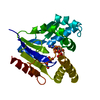
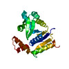
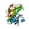
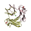
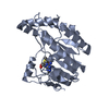
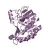
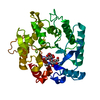
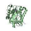
| Title | CMP KINASE FROM ESCHERICHIA COLI FREE ENZYME STRUCTURE | ||||||
 Components Components | PROTEIN (CYTIDINE MONOPHOSPHATE KINASE) | ||||||
 Keywords Keywords | TRANSFERASE / NUCLEOTIDE MONOPHOSPHATE KINASE | ||||||
| Function / homology |  Function and homology information Function and homology information(d)CMP kinase / CMP kinase activity / dCMP kinase activity / pyrimidine nucleotide metabolic process / nucleobase-containing small molecule interconversion / guanosine tetraphosphate binding / response to X-ray / ATP binding / cytosol Similarity search - Function | ||||||
| Biological species |  | ||||||
| Method |  X-RAY DIFFRACTION / SYNCHROTRON / SIRAS / Resolution: 1.75 Å X-RAY DIFFRACTION / SYNCHROTRON / SIRAS / Resolution: 1.75 Å | ||||||
 Authors Authors | Briozzo, P. / Golinelli-Pimpaneau, B. | ||||||
 Citation Citation | Journal: Structure / Year: 1998 Title: Structures of escherichia coli CMP kinase alone and in complex with CDP: a new fold of the nucleoside monophosphate binding domain and insights into cytosine nucleotide specificity. Authors: Briozzo, P. / Golinelli-Pimpaneau, B. / Gilles, A.M. / Gaucher, J.F. / Burlacu-Miron, S. / Sakamoto, H. / Janin, J. / Barzu, O. #1: Journal: J.Biol.Chem. / Year: 1996Title: Cmp Kinase from Escherichia Coli is Structurally Related to Other Nucleoside Monophosphate Kinases Authors: Bucurenci, N. / Sakamoto, H. / Briozzo, P. / Palibroda, N. / Serina, L. / Sarfati, R.S. / Labesse, G. / Briand, G. / Danchin, A. / Barzu, O. / Gilles, A.M. #2: Journal: J.Bacteriol. / Year: 1995Title: The Cmk Gene Encoding Cytidine Monophosphate Kinase is Located in the rspA Operon and is Required for Normal Replication Rate in Escherichia Coli Authors: Fricke, J. / Neuhard, J. / Kelln, R.A. / Pedersen, S. | ||||||
| History |
|
- Structure visualization
| Structure viewer | Molecule: MolmilJmol/JSmol |
|---|
- Downloads & links
-Download
| PDBx/mmCIF format | 1cke.cif.gz | 56.1 KB | Display | PDBx/mmCIF format |
|---|---|---|---|---|
| PDB format | pdb1cke.ent.gz | 40.5 KB | Display | PDB format |
| PDBx/mmJSON format | 1cke.json.gz | Tree view | PDBx/mmJSON format | |
| Others |  Other downloads Other downloads |
-Validation report
| Arichive directory | https://data.pdbj.org/pub/pdb/validation_reports/ck/1ckeftp://data.pdbj.org/pub/pdb/validation_reports/ck/1cke | HTTPS FTP |
|---|
-Related structure data


-Links
 PDBj
PDBj- Assembly
| Deposited unit | 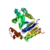
| ||||||||
|---|---|---|---|---|---|---|---|---|---|
| 1 |
| ||||||||
| Unit cell |
|
-Components
| #1: Protein | Mass: 24748.352 Da / Num. of mol.: 1 Source method: isolated from a genetically manipulated source Source: (gene. exp.) |
|---|---|
| #2: Chemical | ChemComp-SO4 /   Mass: 96.063 Da / Num. of mol.: 1 / Source method: obtained synthetically / Formula: SO4 Mass: 96.063 Da / Num. of mol.: 1 / Source method: obtained synthetically / Formula: SO4 |
| #3: Water | ChemComp-HOH /  Mass: 18.015 Da / Num. of mol.: 156 / Source method: isolated from a natural source / Formula: H2O Mass: 18.015 Da / Num. of mol.: 156 / Source method: isolated from a natural source / Formula: H2O |
-Experimental details
-Experiment
| Experiment | Method: X-RAY DIFFRACTION / Number of used crystals: 1 |
|---|
- Sample preparation
| Crystal | Density Matthews: 2.44 Å3/Da / Density % sol: 49 % | ||||||||||||||||||
|---|---|---|---|---|---|---|---|---|---|---|---|---|---|---|---|---|---|---|---|
| Crystal grow | pH: 7.4 / Details: pH 7.4 | ||||||||||||||||||
| Crystal grow | *PLUS Temperature: 20 ℃ / Method: vapor diffusion, hanging drop | ||||||||||||||||||
| Components of the solutions | *PLUS
|
-Data collection
| Diffraction | Mean temperature: 290 K |
|---|---|
| Diffraction source | Source: SYNCHROTRON / Site: LURE  / Beamline: DW32 / Wavelength: 0.97 / Beamline: DW32 / Wavelength: 0.97 |
| Detector | Type: MARRESEARCH / Detector: IMAGE PLATE / Date: Oct 7, 1996 |
| Radiation | Monochromator: GRAPHITE / Protocol: SINGLE WAVELENGTH / Monochromatic (M) / Laue (L): M / Scattering type: x-ray |
| Radiation wavelength | Wavelength: 0.97 Å / Relative weight: 1 |
| Reflection | Resolution: 1.75→30 Å / Num. obs: 23804 / % possible obs: 98.8 % / Observed criterion σ(I): 0 / Redundancy: 7.8 % / Rsym value: 0.06 / Net I/σ(I): 22.9 |
| Reflection shell | Resolution: 1.75→1.81 Å / Mean I/σ(I) obs: 3.6 / Rsym value: 0.395 / % possible all: 99.6 |
| Reflection | *PLUS Num. measured all: 185661 / Rmerge(I) obs: 0.06 |
| Reflection shell | *PLUS % possible obs: 99.6 % / Rmerge(I) obs: 0.395 |
- Processing
| Software |
| ||||||||||||||||||||||||||||||||||||||||||||||||||||||||||||
|---|---|---|---|---|---|---|---|---|---|---|---|---|---|---|---|---|---|---|---|---|---|---|---|---|---|---|---|---|---|---|---|---|---|---|---|---|---|---|---|---|---|---|---|---|---|---|---|---|---|---|---|---|---|---|---|---|---|---|---|---|---|
| Refinement | Method to determine structure: SIRAS / Resolution: 1.75→7 Å / σ(F): 2 Details: SIDE CHAIN FROM RESIDUE ARG 173 WAS NOT WELL DEFINED IN THE DENSITY AND WAS MODELED BY STEREOCHEMISTRY
| ||||||||||||||||||||||||||||||||||||||||||||||||||||||||||||
| Displacement parameters | Biso mean: 33.6 Å2 | ||||||||||||||||||||||||||||||||||||||||||||||||||||||||||||
| Refinement step | Cycle: LAST / Resolution: 1.75→7 Å
| ||||||||||||||||||||||||||||||||||||||||||||||||||||||||||||
| Refine LS restraints |
| ||||||||||||||||||||||||||||||||||||||||||||||||||||||||||||
| Software | *PLUS Name: X-PLOR / Version: 3.84 / Classification: refinement | ||||||||||||||||||||||||||||||||||||||||||||||||||||||||||||
| Refinement | *PLUS Lowest resolution: 7 Å / σ(F): 2 / % reflection Rfree: 10 % | ||||||||||||||||||||||||||||||||||||||||||||||||||||||||||||
| Solvent computation | *PLUS | ||||||||||||||||||||||||||||||||||||||||||||||||||||||||||||
| Displacement parameters | *PLUS Biso mean: 33.6 Å2 | ||||||||||||||||||||||||||||||||||||||||||||||||||||||||||||
| Refine LS restraints | *PLUS
|
