Mass: 18.015 Da / Num. of mol.: 12 / Source method: isolated from a natural source / Formula: H2O
Sequence details
NO ELECTRON DENSITY WAS OBSERVED FOR THE FIRST 13 RESIDUES. IN CASES WHERE NO SIDECHAIN ELECTRON ...NO ELECTRON DENSITY WAS OBSERVED FOR THE FIRST 13 RESIDUES. IN CASES WHERE NO SIDECHAIN ELECTRON DENSITY WAS OBSERVED, RESIDUES WERE MODELED AS ALA DURING THE REFINEMENT AND ITERATIVE REBUILDING. IN ACCORDANCE WITH RCSB DIRECTIVES, SUCH RESIDUES HAVE BEEN ASSIGNED RESIDUE TYPES BASED ON THE EXPECTED SEQUENCE FOR RABBIT LIVER CYTOSILIC SHMT (SWS ENTRY P07511); HOWEVER COORDINATES ARE NOT INCLUDED FOR SIDECHAIN ATOMS BEYOND CB. THESE RESIDUES INCLUDE: MODEL SEQUENCE SWS P07511 SEQUENCE HIS A -5 HIS 17 LYS A 7 LYS 26 LYS A 20 LYS 37 GLN A 62 GLN 79 GLU A 69 GLU 86 LYS A 80 LYS 97 GLN A 892 GLN 108 LYS A 1341 LYS 156 LYS A 1342 LYS 157 LYS A 277 LYS 316 LYS A 354 LYS 392 SER A 355 SER 393 LYS A 375 LYS 415 PRO A 3962 PRO 440 ARG A 3963 ARG 441 LYS A 3991 LYS 445 LYS A 3994 LYS 448 LYS A 401 LYS 450 LYS A 405 LYS 456 ARG A 407 ARG 459 ARG A 410 ARG 462 GLN A 413 GLN 466 LEU A 4181 LEU 474 TRP B -8 TRP 14 HIS B -5 HIS 17 LYS B 7 LYS 26 LYS B 20 LYS 37 ARG B 41 ARG 58 LYS B 1341 LYS 156 LYS B 168 LYS 195 VAL B 2441 VAL 272 SER B 2443 SER 274 VAL B 2444 VAL 275 LYS B 245 LYS 281 ILE B 283 ILE 283 LYS B 354 LYS 393 SER B 355 SER 394 LEU B 357 LEU 396 ARG B 358 ARG 397 LYS B 380 LYS 419 ARG B 3963 ARG 441 LYS B 3991 LYS 445 LYS B 3994 LYS 448 GLU B 400 GLU 449 LYS B 401 LYS 450 LYS B 405 LYS 456 ARG B 407 ARG 459 GLN B 413 GLN 466
-
Experimental details
-
Experiment
Experiment
Method: X-RAY DIFFRACTION / Number of used crystals: 5
-
Sample preparation
Crystal
Density Matthews: 2.85 Å3/Da / Density % sol: 57 %
Crystal grow
Method: vapor diffusion, hanging drop / pH: 6 Details: 20MM K2HPO4, KH2PO4, 25MM POTASIUM MES OR SODIUM HEPES, PH 7.0 0.2 MM 2-MERCAPTOETHANOL, 3% PEG 400, pH 6.0, VAPOR DIFFUSION, HANGING DROP
Monochromator: GRAPHITE / Protocol: SINGLE WAVELENGTH / Monochromatic (M) / Laue (L): M / Scattering type: x-ray
Radiation wavelength
Wavelength: 1.5418 Å / Relative weight: 1
Reflection
Resolution: 2.8→35 Å / Num. obs: 28327 / % possible obs: 100 % / Observed criterion σ(I): -3 / Redundancy: 19 % / Biso Wilson estimate: 88.8 Å2 / Rmerge(I) obs: 0.173 / Net I/σ(I): 10.9
Reflection shell
*PLUS
Mean I/σ(I) obs: 1
-
Processing
Software
Name
Version
Classification
PHASES
phasing
CNS
0.5
refinement
DENZO
datareduction
SCALEPACK
datascaling
Refinement
Method to determine structure: MIRAS / Resolution: 2.8→20 Å / Rfactor Rfree error: 0.005 / Data cutoff high rms absF: 2580178.46 / Isotropic thermal model: GROUP / Cross valid method: THROUGHOUT / σ(F): 0 Details: PROTEIN+PLP REFINED WITH TORSION ANGLE DYNAMICS, WATERS HELD FIXED. WATERS REFINED WITH CARTESIAN DYNAMICS, PROTEIN + PLP HELD FIXED. POSITIONAL NCS RESTRAINTS APPLIED BETWEEN EQUIVALENT ...Details: PROTEIN+PLP REFINED WITH TORSION ANGLE DYNAMICS, WATERS HELD FIXED. WATERS REFINED WITH CARTESIAN DYNAMICS, PROTEIN + PLP HELD FIXED. POSITIONAL NCS RESTRAINTS APPLIED BETWEEN EQUIVALENT RESIDUES IN CHAINS A + B. RESIDUES 9992- 1, 244-247, 4181-4180 AND 2291 WERE NOT RESTRAINED
In the structure databanks used in Yorodumi, some data are registered as the other names, "COVID-19 virus" and "2019-nCoV". Here are the details of the virus and the list of structure data.
Jan 31, 2019. EMDB accession codes are about to change! (news from PDBe EMDB page)
EMDB accession codes are about to change! (news from PDBe EMDB page)
The allocation of 4 digits for EMDB accession codes will soon come to an end. Whilst these codes will remain in use, new EMDB accession codes will include an additional digit and will expand incrementally as the available range of codes is exhausted. The current 4-digit format prefixed with “EMD-” (i.e. EMD-XXXX) will advance to a 5-digit format (i.e. EMD-XXXXX), and so on. It is currently estimated that the 4-digit codes will be depleted around Spring 2019, at which point the 5-digit format will come into force.
The EM Navigator/Yorodumi systems omit the EMD- prefix.
Related info.:Q: What is EMD? / ID/Accession-code notation in Yorodumi/EM Navigator
Yorodumi is a browser for structure data from EMDB, PDB, SASBDB, etc.
This page is also the successor to EM Navigator detail page, and also detail information page/front-end page for Omokage search.
The word "yorodu" (or yorozu) is an old Japanese word meaning "ten thousand". "mi" (miru) is to see.
Related info.:EMDB / PDB / SASBDB / Comparison of 3 databanks / Yorodumi Search / Aug 31, 2016. New EM Navigator & Yorodumi / Yorodumi Papers / Jmol/JSmol / Function and homology information / Changes in new EM Navigator and Yorodumi
 Movie
Movie Controller

 Yorodumi
 Open data
 Basic information
 Components
 Keywords
 Function and homology information
 X-RAY DIFFRACTION /
  Authors
 Citation
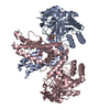
 Structure visualization
 Downloads & links
 Other downloads


 PDBj
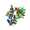
PDBj Assembly


 Mass: 247.142 Da / Num. of mol.: 2 / Source method: obtained synthetically / Formula: C8H10NO6P
 Mass: 18.015 Da / Num. of mol.: 12 / Source method: isolated from a natural source / Formula: H2O
 Sample preparation
 Processing