 Movie
Movie Controller

[English] 日本語
 Yorodumi
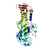
- PDB-1cdd: STRUCTURES OF APO AND COMPLEXED ESCHERICHIA COLI GLYCINAMIDE RIBO... -
+ Open data

- Basic information
| Entry | Database: PDB / ID: 1cdd | ||||||
|---|---|---|---|---|---|---|---|
| Title | STRUCTURES OF APO AND COMPLEXED ESCHERICHIA COLI GLYCINAMIDE RIBONUCLEOTIDE TRANSFORMYLASE | ||||||
 Components Components | PHOSPHORIBOSYL-GLYCINAMIDE FORMYLTRANSFERASE | ||||||
 Keywords Keywords | TRANSFERASE(FORMYL) | ||||||
| Function / homology |  Function and homology information Function and homology informationphosphoribosylglycinamide formyltransferase 1 / phosphoribosylglycinamide formyltransferase activity / 'de novo' IMP biosynthetic process / DNA damage response / cytoplasm / cytosol Similarity search - Function | ||||||
| Biological species |  | ||||||
| Method |  X-RAY DIFFRACTION / Resolution: 2.8 Å X-RAY DIFFRACTION / Resolution: 2.8 Å | ||||||
 Authors Authors | Almassy, R.J. / Janson, C.A. / Kan, C.-C. / Hostomska, Z. | ||||||
 Citation Citation | Journal: Proc.Natl.Acad.Sci.USA / Year: 1992 Title: Structures of apo and complexed Escherichia coli glycinamide ribonucleotide transformylase. Authors: Almassy, R.J. / Janson, C.A. / Kan, C.C. / Hostomska, Z. #1: Journal: Nucleic Acids Res. / Year: 1990Title: De Novo Purine Nucleotide Biosynthesis: Cloning of Human and Avian Cdna'S Encoding the Trifunctional Glycinamide Ribonucleotide Synthetase-Aminoimidazole Ribonucleotide Synthetase-Glycinamide ...Title: De Novo Purine Nucleotide Biosynthesis: Cloning of Human and Avian Cdna'S Encoding the Trifunctional Glycinamide Ribonucleotide Synthetase-Aminoimidazole Ribonucleotide Synthetase-Glycinamide Ribonucleotide Transformylase by Functional Complementation in E. Coli Authors: Aimi, J. / Qiu, H. / Williams, J. / Zalkin, H. / Dixon, J.E. #2: Journal: J.Biol.Chem. / Year: 1987Title: Identification and Nucleotide Sequence of a Gene Encoding 5'-Phosphoribosylglycinamide Transformylase in Escherichia Coli K12 Authors: Smith, J.M. / Daum III, H.A. | ||||||
| History |
|
- Structure visualization
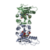
| Structure viewer | Molecule: MolmilJmol/JSmol |
|---|
- Downloads & links
-Download
| PDBx/mmCIF format | 1cdd.cif.gz | 84.4 KB | Display | PDBx/mmCIF format |
|---|---|---|---|---|
| PDB format | pdb1cdd.ent.gz | 65 KB | Display | PDB format |
| PDBx/mmJSON format | 1cdd.json.gz | Tree view | PDBx/mmJSON format | |
| Others |  Other downloads Other downloads |
-Validation report
| Arichive directory | https://data.pdbj.org/pub/pdb/validation_reports/cd/1cddftp://data.pdbj.org/pub/pdb/validation_reports/cd/1cdd | HTTPS FTP |
|---|
-Related structure data


-Links
 PDBj
PDBj- Assembly
| Deposited unit | 
| ||||||||
|---|---|---|---|---|---|---|---|---|---|
| 1 |
| ||||||||
| 2 | 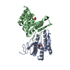
| ||||||||
| Unit cell |
| ||||||||
| Atom site foot note | 1: PRO B 204 - PRO B 205 OMEGA =212.30 PEPTIDE BOND DEVIATES SIGNIFICANTLY FROM TRANS CONFORMATION | ||||||||
| Noncrystallographic symmetry (NCS) | NCS oper: (Code: given Matrix: (-0.19391, 0.33875, 0.92068), Vector: Details | THE TRANSFORMATION PRESENTED ON *MTRIX* RECORDS BELOW WILL YIELD APPROXIMATE COORDINATES FOR MOLECULE 1 (CHAIN A) WHEN APPLIED TO MOLECULE 2 (CHAIN B) WITH AN RMS DEVIATION OF 0.83 ANGSTROMS FOR 189 CA COORDINATES (APPROXIMATELY A TWO-FOLD ROTATION, 184 DEGREES). | |
-Components
| #1: Protein | Mass: 23266.254 Da / Num. of mol.: 2 Source method: isolated from a genetically manipulated source Source: (gene. exp.) References: UniProt: P08179, phosphoribosylglycinamide formyltransferase 1 #2: Chemical |   Mass: 94.971 Da / Num. of mol.: 2 / Source method: obtained synthetically / Formula: PO4 Mass: 94.971 Da / Num. of mol.: 2 / Source method: obtained synthetically / Formula: PO4 |
|---|
-Experimental details
-Experiment
| Experiment | Method: X-RAY DIFFRACTION |
|---|
- Sample preparation
| Crystal | Density Matthews: 3.78 Å3/Da / Density % sol: 67.47 % | |||||||||||||||||||||||||
|---|---|---|---|---|---|---|---|---|---|---|---|---|---|---|---|---|---|---|---|---|---|---|---|---|---|---|
| Crystal grow | *PLUS Temperature: 20 ℃ / pH: 7.5 / Method: vapor diffusion, hanging drop | |||||||||||||||||||||||||
| Components of the solutions | *PLUS
|
-Data collection
| Radiation | Scattering type: x-ray |
|---|---|
| Radiation wavelength | Relative weight: 1 |
| Reflection | *PLUS Highest resolution: 2.8 Å / Lowest resolution: 8 Å / Num. all: 124304 / Num. obs: 16623 / Rmerge(I) obs: 0.074 |
- Processing
| Software |
| |||||||||||||||||||||||||||||||||||||||||||||||||||||||||||||||
|---|---|---|---|---|---|---|---|---|---|---|---|---|---|---|---|---|---|---|---|---|---|---|---|---|---|---|---|---|---|---|---|---|---|---|---|---|---|---|---|---|---|---|---|---|---|---|---|---|---|---|---|---|---|---|---|---|---|---|---|---|---|---|---|---|
| Refinement | Rfactor obs: 0.225 / Highest resolution: 2.8 Å Details: RESIDUES 210 - 212 IN CHAIN A HAVE HIGH TEMPERATURE FACTORS, ARE IN WEAK DENSITY, AND HAVE SIGNIFICANT DISORDER. | |||||||||||||||||||||||||||||||||||||||||||||||||||||||||||||||
| Refinement step | Cycle: LAST / Highest resolution: 2.8 Å
| |||||||||||||||||||||||||||||||||||||||||||||||||||||||||||||||
| Refine LS restraints |
| |||||||||||||||||||||||||||||||||||||||||||||||||||||||||||||||
| Refinement | *PLUS Highest resolution: 2.8 Å / Lowest resolution: 8 Å / Num. reflection obs: 15431 / σ(F): 0.5 / Rfactor obs: 0.225 | |||||||||||||||||||||||||||||||||||||||||||||||||||||||||||||||
| Solvent computation | *PLUS | |||||||||||||||||||||||||||||||||||||||||||||||||||||||||||||||
| Displacement parameters | *PLUS | |||||||||||||||||||||||||||||||||||||||||||||||||||||||||||||||
| Refine LS restraints | *PLUS
|
