 Movie
Movie Controller


+ Open data

- Basic information
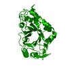
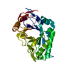
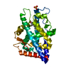
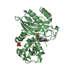
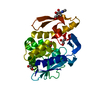
| Entry | Database: PDB / ID: 1c90 | ||||||
|---|---|---|---|---|---|---|---|
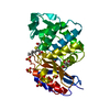
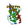
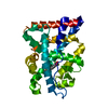
| Title | Endo-Beta-N-Acetylglucosaminidase H, E132Q Mutant | ||||||
 Components Components | ENDO-BETA-N-ACETYLGLUCOSAMINIDASE H | ||||||
 Keywords Keywords | HYDROLASE / (BETA/ALPHA)8 BARREL | ||||||
| Function / homology |  Function and homology information Function and homology informationmannosyl-glycoprotein endo-beta-N-acetylglucosaminidase / mannosyl-glycoprotein endo-beta-N-acetylglucosaminidase activity / carbohydrate metabolic process / extracellular region Similarity search - Function | ||||||
| Biological species |  Streptomyces plicatus (bacteria) Streptomyces plicatus (bacteria) | ||||||
| Method |  X-RAY DIFFRACTION / Resolution: 2.1 Å X-RAY DIFFRACTION / Resolution: 2.1 Å | ||||||
 Authors Authors | Rao, V. / Tao, C. / Guan, C. / Van Roey, P. | ||||||
 Citation Citation | Journal: Protein Sci. / Year: 1999 Title: Mutations of endo-beta-N-acetylglucosaminidase H active site residues Assp130 and Glu132: activities and conformations. Authors: Rao, V. / Cui, T. / Guan, C. / Van Roey, P. | ||||||
| History |
|
- Structure visualization
| Structure viewer | Molecule: MolmilJmol/JSmol |
|---|
- Downloads & links
-Download
| PDBx/mmCIF format | 1c90.cif.gz | 115.1 KB | Display | PDBx/mmCIF format |
|---|---|---|---|---|
| PDB format | pdb1c90.ent.gz | 89 KB | Display | PDB format |
| PDBx/mmJSON format | 1c90.json.gz | Tree view | PDBx/mmJSON format | |
| Others |  Other downloads Other downloads |
-Validation report
| Arichive directory | https://data.pdbj.org/pub/pdb/validation_reports/c9/1c90ftp://data.pdbj.org/pub/pdb/validation_reports/c9/1c90 | HTTPS FTP |
|---|
-Related structure data
| Related structure data |  1c3fC 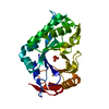 1c8xC  1c8yC  1c91C  1c92C  1c93C C: citing same article ( |
|---|---|
| Similar structure data |


-Links
 PDBj
PDBj- Assembly
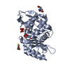
| Deposited unit | 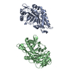
| ||||||||
|---|---|---|---|---|---|---|---|---|---|
| 1 | 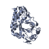
| ||||||||
| 2 | 
| ||||||||
| Unit cell |
|
-Components
| #1: Protein | Mass: 28524.428 Da / Num. of mol.: 2 / Mutation: E132Q Source method: isolated from a genetically manipulated source Source: (gene. exp.) Streptomyces plicatus (bacteria) / Plasmid: PMAL / Production host: References: UniProt: P04067, mannosyl-glycoprotein endo-beta-N-acetylglucosaminidase #2: Water | ChemComp-HOH / |  Mass: 18.015 Da / Num. of mol.: 319 / Source method: isolated from a natural source / Formula: H2O Mass: 18.015 Da / Num. of mol.: 319 / Source method: isolated from a natural source / Formula: H2O |
|---|
-Experimental details
-Experiment
| Experiment | Method: X-RAY DIFFRACTION / Number of used crystals: 1 |
|---|
- Sample preparation
| Crystal | Density Matthews: 2.04 Å3/Da / Density % sol: 39.76 % | ||||||||||||||||||||||||||||||||||||||||||||||||||||||||
|---|---|---|---|---|---|---|---|---|---|---|---|---|---|---|---|---|---|---|---|---|---|---|---|---|---|---|---|---|---|---|---|---|---|---|---|---|---|---|---|---|---|---|---|---|---|---|---|---|---|---|---|---|---|---|---|---|---|
| Crystal grow | Temperature: 293 K / Method: vapor diffusion, hanging drop / pH: 6.5 Details: 19% PEG1000, 100 MM ZN(AC)2 100 MM CACODYLATE, pH 6.5, VAPOR DIFFUSION, HANGING DROP, temperature 293K | ||||||||||||||||||||||||||||||||||||||||||||||||||||||||
| Crystal grow | *PLUS | ||||||||||||||||||||||||||||||||||||||||||||||||||||||||
| Components of the solutions | *PLUS
|
-Data collection
| Diffraction | Mean temperature: 100 K |
|---|---|
| Diffraction source | Source: ROTATING ANODE / Type: RIGAKU RU200 / Wavelength: 1.5418 |
| Detector | Type: RIGAKU RAXIS IIC / Detector: IMAGE PLATE / Date: Jan 1, 1996 |
| Radiation | Protocol: SINGLE WAVELENGTH / Monochromatic (M) / Laue (L): M / Scattering type: x-ray |
| Radiation wavelength | Wavelength: 1.5418 Å / Relative weight: 1 |
| Reflection | Resolution: 2.1→20 Å / Num. all: 51615 / Num. obs: 27256 / % possible obs: 87.6 % / Observed criterion σ(F): 2 / Rmerge(I) obs: 0.066 |
| Reflection shell | Resolution: 2.1→2.2 Å / % possible all: 83 |
| Reflection | *PLUS % possible obs: 91 % / Redundancy: 3 % / Rmerge(I) obs: 0.076 |
| Reflection shell | *PLUS % possible obs: 88 % |
- Processing
| Software |
| ||||||||||||||||||||||||||||||||||||||||||||||||||||||||||||
|---|---|---|---|---|---|---|---|---|---|---|---|---|---|---|---|---|---|---|---|---|---|---|---|---|---|---|---|---|---|---|---|---|---|---|---|---|---|---|---|---|---|---|---|---|---|---|---|---|---|---|---|---|---|---|---|---|---|---|---|---|---|
| Refinement | Resolution: 2.1→10 Å / σ(F): 2
| ||||||||||||||||||||||||||||||||||||||||||||||||||||||||||||
| Refinement step | Cycle: LAST / Resolution: 2.1→10 Å
| ||||||||||||||||||||||||||||||||||||||||||||||||||||||||||||
| Refine LS restraints |
| ||||||||||||||||||||||||||||||||||||||||||||||||||||||||||||
| Software | *PLUS Name: X-PLOR / Version: 3.1 / Classification: refinement | ||||||||||||||||||||||||||||||||||||||||||||||||||||||||||||
| Refinement | *PLUS Highest resolution: 2.1 Å / Lowest resolution: 10 Å / σ(F): 2 / Rfactor obs: 0.212 / Rfactor Rwork: 0.2 | ||||||||||||||||||||||||||||||||||||||||||||||||||||||||||||
| Solvent computation | *PLUS | ||||||||||||||||||||||||||||||||||||||||||||||||||||||||||||
| Displacement parameters | *PLUS | ||||||||||||||||||||||||||||||||||||||||||||||||||||||||||||
| Refine LS restraints | *PLUS
|
