 Movie
Movie Controller


+ Open data

- Basic information
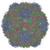
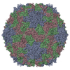
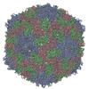
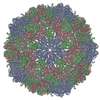
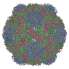
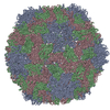
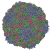
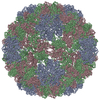
| Entry | Database: PDB / ID: 1c8n | ||||||
|---|---|---|---|---|---|---|---|
| Title | TOBACCO NECROSIS VIRUS | ||||||
 Components Components | COAT PROTEIN | ||||||
 Keywords Keywords | VIRUS / Icosahedral virus | ||||||
| Function / homology |  Function and homology information Function and homology informationT=3 icosahedral viral capsid / structural molecule activity / metal ion binding Similarity search - Function | ||||||
| Biological species |  unidentified tobacco necrosis virus unidentified tobacco necrosis virus | ||||||
| Method |  X-RAY DIFFRACTION / SYNCHROTRON / MOLECULAR REPLACEMENT / Resolution: 2.25 Å X-RAY DIFFRACTION / SYNCHROTRON / MOLECULAR REPLACEMENT / Resolution: 2.25 Å | ||||||
 Authors Authors | Oda, Y. / Fukuyama, K. | ||||||
 Citation Citation | Journal: J.Mol.Biol. / Year: 2000 Title: Crystal structure of tobacco necrosis virus at 2.25 A resolution. Authors: Oda, Y. / Saeki, K. / Takahashi, Y. / Maeda, T. / Naitow, H. / Tsukihara, T. / Fukuyama, K. #1: Journal: Acta Crystallogr.,Sect.D / Year: 1994Title: Crystal Structural Analysis of Tobacco Necrosis Virus at 5 Angstrom Resolution Authors: Bando, M. / Morimoto, Y. / Sato, T. / Tsukihara, T. / Yokota, Y. / Fukuyama, K. / Matsubara, H. #2: Journal: Acta Crystallogr.,Sect.B / Year: 1990Title: Symmetry and Subunit Arrangement of Tobacco Necrosis Virus (TNV) Authors: Tsukihara, T. / Yokota, Y. / Koyama, T. / Fukuyama, K. / Matsubara, H. #3: Journal: J.Mol.Biol. / Year: 1987Title: Crystallization and Preliminary X-Ray Diffraction Studies of Tobacco Necrosis Virus Authors: Fukuyama, K. / Hirota, S. / Tsukihara, T. | ||||||
| History |
|
- Structure visualization
| Structure viewer | Molecule: MolmilJmol/JSmol |
|---|
- Downloads & links
-Download
| PDBx/mmCIF format | 1c8n.cif.gz | 130.4 KB | Display | PDBx/mmCIF format |
|---|---|---|---|---|
| PDB format | pdb1c8n.ent.gz | 101 KB | Display | PDB format |
| PDBx/mmJSON format | 1c8n.json.gz | Tree view | PDBx/mmJSON format | |
| Others |  Other downloads Other downloads |
-Validation report
| Arichive directory | https://data.pdbj.org/pub/pdb/validation_reports/c8/1c8nftp://data.pdbj.org/pub/pdb/validation_reports/c8/1c8n | HTTPS FTP |
|---|
-Related structure data
| Related structure data | 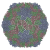 1tnvS S: Starting model for refinement |
|---|---|
| Similar structure data |

-Links
 PDBj
PDBj

- Assembly
| Deposited unit | 
| ||||||||||||||||||
|---|---|---|---|---|---|---|---|---|---|---|---|---|---|---|---|---|---|---|---|
| 1 | x 60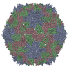
| ||||||||||||||||||
| 2 |
| ||||||||||||||||||
| 3 | x 5
| ||||||||||||||||||
| 4 | x 6
| ||||||||||||||||||
| 5 | 
| ||||||||||||||||||
| 6 | x 5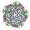
| ||||||||||||||||||
| Unit cell |
| ||||||||||||||||||
| Symmetry | Point symmetry: (Hermann–Mauguin notation: 532 / Schoenflies symbol: I (icosahedral)) | ||||||||||||||||||
| Noncrystallographic symmetry (NCS) | NCS oper:
|
-Components
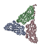
| #1: Protein | Mass: 29985.188 Da / Num. of mol.: 3 / Source method: isolated from a natural source / Details: THIS STRAIN IS CLOSE TO STRAIN A. / Source: (natural) unidentified tobacco necrosis virus / Genus: Necrovirus / Strain: TOYAMA ISOLATE / References: UniProt: Q9IPS9#2: Chemical | ChemComp-CA /   Mass: 40.078 Da / Num. of mol.: 5 / Source method: obtained synthetically / Formula: Ca Mass: 40.078 Da / Num. of mol.: 5 / Source method: obtained synthetically / Formula: Ca#3: Water | ChemComp-HOH / |  Mass: 18.015 Da / Num. of mol.: 192 / Source method: isolated from a natural source / Formula: H2O Mass: 18.015 Da / Num. of mol.: 192 / Source method: isolated from a natural source / Formula: H2O |
|---|
-Experimental details
-Experiment
| Experiment | Method: X-RAY DIFFRACTION / Number of used crystals: 2 |
|---|
- Sample preparation
| Crystal grow | pH: 6 / Details: pH 6.0 |
|---|---|
| Crystal grow | *PLUS Method: microdialysis |
| Components of the solutions | *PLUS Conc.: 0.4 M / Common name: sodium phosphate |
-Data collection
| Diffraction | Mean temperature: 293 K |
|---|---|
| Diffraction source | Source: SYNCHROTRON / Site: Photon Factory  / Beamline: BL-6A / Wavelength: 1 / Beamline: BL-6A / Wavelength: 1 |
| Detector | Type: FUJI / Detector: IMAGE PLATE / Details: MIRRORS |
| Radiation | Protocol: SINGLE WAVELENGTH / Monochromatic (M) / Laue (L): M / Scattering type: x-ray |
| Radiation wavelength | Wavelength: 1 Å / Relative weight: 1 |
| Reflection | Resolution: 2.25→50 Å / Num. obs: 298768 / % possible obs: 99 % / Observed criterion σ(I): 2 / Redundancy: 2.8 % / Rmerge(I) obs: 0.079 / Net I/σ(I): 3.9 |
| Reflection shell | Resolution: 2.25→2.37 Å / Redundancy: 1.9 % / Rmerge(I) obs: 0.392 / Mean I/σ(I) obs: 1.2 / % possible all: 95.6 |
| Reflection | *PLUS % possible obs: 99 % / Num. measured all: 842131 |
| Reflection shell | *PLUS % possible obs: 95.6 % |
- Processing
| Software |
| ||||||||||||||||||
|---|---|---|---|---|---|---|---|---|---|---|---|---|---|---|---|---|---|---|---|
| Refinement | Method to determine structure: MOLECULAR REPLACEMENT Starting model: 1TNV Resolution: 2.25→8 Å / Isotropic thermal model: RESTRAINED / σ(F): 2
| ||||||||||||||||||
| Refinement step | Cycle: LAST / Resolution: 2.25→8 Å
| ||||||||||||||||||
| Refine LS restraints |
| ||||||||||||||||||
| Software | *PLUS Name: X-PLOR / Version: 3 / Classification: refinement | ||||||||||||||||||
| Refinement | *PLUS Lowest resolution: 8 Å / σ(F): 2 | ||||||||||||||||||
| Solvent computation | *PLUS | ||||||||||||||||||
| Displacement parameters | *PLUS |
