 Movie
Movie Controller


+ Open data

- Basic information
| Entry | Database: PDB / ID: 1c5e | ||||||
|---|---|---|---|---|---|---|---|
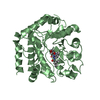
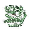
| Title | BACTERIOPHAGE LAMBDA HEAD PROTEIN D | ||||||
 Components Components | HEAD DECORATION PROTEIN | ||||||
 Keywords Keywords | VIRAL PROTEIN / BACTERIOPHAGE LAMBDA / HEAD PROTEIN D / PROTEIN CRYSTAL STRUCTURE / VIRUS ASSEMBLY / PHAGE DISPLAY | ||||||
| Function / homology |  Function and homology information Function and homology informationviral capsid, decoration / viral DNA genome packaging / host cell cytoplasm Similarity search - Function | ||||||
| Biological species |  Enterobacteria phage lambda (virus) Enterobacteria phage lambda (virus) | ||||||
| Method |  X-RAY DIFFRACTION / SYNCHROTRON / MAD / Resolution: 1.1 Å X-RAY DIFFRACTION / SYNCHROTRON / MAD / Resolution: 1.1 Å | ||||||
 Authors Authors | Yang, F. / Forrer, P. / Dauter, Z. / Pluckthun, A. / Wlodawer, A. | ||||||
 Citation Citation | Journal: Nat.Struct.Biol. / Year: 2000 Title: Novel fold and capsid-binding properties of the lambda-phage display platform protein gpD. Authors: Yang, F. / Forrer, P. / Dauter, Z. / Conway, J.F. / Cheng, N. / Cerritelli, M.E. / Steven, A.C. / Pluckthun, A. / Wlodawer, A. | ||||||
| History |
|
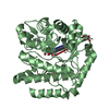
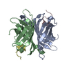
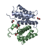
- Structure visualization
| Structure viewer | Molecule: MolmilJmol/JSmol |
|---|
- Downloads & links
-Download
| PDBx/mmCIF format | 1c5e.cif.gz | 127 KB | Display | PDBx/mmCIF format |
|---|---|---|---|---|
| PDB format | pdb1c5e.ent.gz | 100.2 KB | Display | PDB format |
| PDBx/mmJSON format | 1c5e.json.gz | Tree view | PDBx/mmJSON format | |
| Others |  Other downloads Other downloads |
-Validation report
| Arichive directory | https://data.pdbj.org/pub/pdb/validation_reports/c5/1c5eftp://data.pdbj.org/pub/pdb/validation_reports/c5/1c5e | HTTPS FTP |
|---|
-Related structure data
| Similar structure data |
|---|


-Links
 PDBj
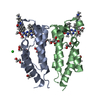
PDBj- Assembly
| Deposited unit | 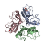
| ||||||||||||
|---|---|---|---|---|---|---|---|---|---|---|---|---|---|
| 1 |
| ||||||||||||
| Unit cell |
| ||||||||||||
| Noncrystallographic symmetry (NCS) | NCS oper:
|
-Components
| #1: Protein | Mass: 9829.972 Da / Num. of mol.: 3 Source method: isolated from a genetically manipulated source Source: (gene. exp.) Enterobacteria phage lambda (virus) / Genus: Lambda-like viruses / References: UniProt: P03712#2: Chemical |   Mass: 92.094 Da / Num. of mol.: 2 / Source method: obtained synthetically / Formula: C3H8O3 Mass: 92.094 Da / Num. of mol.: 2 / Source method: obtained synthetically / Formula: C3H8O3#3: Water | ChemComp-HOH / |  Mass: 18.015 Da / Num. of mol.: 309 / Source method: isolated from a natural source / Formula: H2O Mass: 18.015 Da / Num. of mol.: 309 / Source method: isolated from a natural source / Formula: H2O |
|---|
-Experimental details
-Experiment
| Experiment | Method: X-RAY DIFFRACTION / Number of used crystals: 1 |
|---|
- Sample preparation
| Crystal | Density Matthews: 2 Å3/Da / Density % sol: 39 % | ||||||||||||||||||||||||||||||
|---|---|---|---|---|---|---|---|---|---|---|---|---|---|---|---|---|---|---|---|---|---|---|---|---|---|---|---|---|---|---|---|
| Crystal grow | pH: 6.5 / Details: 28% PEG 4000, 0.1 M BIS-TRIS PH 6.5, 10 % GLYCEROL | ||||||||||||||||||||||||||||||
| Crystal grow | *PLUS pH: 7.5 / Method: vapor diffusion, hanging drop | ||||||||||||||||||||||||||||||
| Components of the solutions | *PLUS
|
-Data collection
| Diffraction | Mean temperature: 100 K |
|---|---|
| Diffraction source | Source: SYNCHROTRON / Site: NSLS  / Beamline: X9B / Wavelength: 0.98 / Beamline: X9B / Wavelength: 0.98 |
| Detector | Type: MAR scanner 345 mm plate / Detector: IMAGE PLATE / Date: Apr 1, 1999 |
| Radiation | Protocol: SINGLE WAVELENGTH / Monochromatic (M) / Laue (L): M / Scattering type: x-ray |
| Radiation wavelength | Wavelength: 0.98 Å / Relative weight: 1 |
| Reflection | Resolution: 1.1→20 Å / Num. obs: 109996 / % possible obs: 99.2 % / Observed criterion σ(I): -3 / Redundancy: 5.8 % / Rsym value: 4.6 |
| Reflection shell | Resolution: 1.1→1.12 Å / Rsym value: 25.6 / % possible all: 98.3 |
| Reflection | *PLUS Highest resolution: 1.1 Å / Lowest resolution: 20 Å / Num. measured all: 635194 / Rmerge(I) obs: 0.046 |
- Processing
| Software |
| |||||||||||||||||||||||||||||||||
|---|---|---|---|---|---|---|---|---|---|---|---|---|---|---|---|---|---|---|---|---|---|---|---|---|---|---|---|---|---|---|---|---|---|---|
| Refinement | Method to determine structure: MAD / Resolution: 1.1→10 Å / Num. parameters: 21691 / Num. restraintsaints: 26304 / Cross valid method: THROUGHOUT / σ(F): 0 / Stereochemistry target values: ENGH AND HUBER / Details: ANISOTROPIC REFINEMENT REDUCED FREE R (NO CUTOFF)
| |||||||||||||||||||||||||||||||||
| Solvent computation | Solvent model: MOEWS & KRETSINGER, J.MOL.BIOL.91(1973)201-228 | |||||||||||||||||||||||||||||||||
| Refine analyze | Num. disordered residues: 6 / Occupancy sum hydrogen: 2051 / Occupancy sum non hydrogen: 2394 | |||||||||||||||||||||||||||||||||
| Refinement step | Cycle: LAST / Resolution: 1.1→10 Å
| |||||||||||||||||||||||||||||||||
| Refine LS restraints |
| |||||||||||||||||||||||||||||||||
| Software | *PLUS Name: SHELXL-97 / Classification: refinement | |||||||||||||||||||||||||||||||||
| Refinement | *PLUS Lowest resolution: 10 Å / σ(F): 0 / % reflection Rfree: 5.1 % / Rfactor obs: 0.0982 / Rfactor Rwork: 0.099 | |||||||||||||||||||||||||||||||||
| Solvent computation | *PLUS | |||||||||||||||||||||||||||||||||
| Displacement parameters | *PLUS | |||||||||||||||||||||||||||||||||
| Refine LS restraints | *PLUS Type: s_chiral_restr / Dev ideal: 0.087 |
