 Movie
Movie Controller


+ Open data

- Basic information
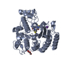
| Entry | Database: PDB / ID: 1c3s | ||||||
|---|---|---|---|---|---|---|---|
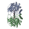
| Title | CRYSTAL STRUCTURE OF AN HDAC HOMOLOG COMPLEXED WITH SAHA | ||||||
 Components Components | HDLP (HISTONE DEACETYLASE-LIKE PROTEIN) | ||||||
 Keywords Keywords | LYASE / ALPHA/BETA FOLD / HYDROXAMIC ACID / PENTA-COORDINATED ZINC / CHARGE-RELAY SYSTEM | ||||||
| Function / homology |  Function and homology information Function and homology informationacetoin catabolic process / histone deacetylase activity / epigenetic regulation of gene expression / metal ion binding Similarity search - Function | ||||||
| Biological species |   Aquifex aeolicus (bacteria) Aquifex aeolicus (bacteria) | ||||||
| Method |  X-RAY DIFFRACTION / Resolution: 2.5 Å X-RAY DIFFRACTION / Resolution: 2.5 Å | ||||||
 Authors Authors | Finnin, M.S. / Donigian, J.R. / Pavletich, N.P. | ||||||
 Citation Citation | Journal: Nature / Year: 1999 Title: Structures of a histone deacetylase homologue bound to the TSA and SAHA inhibitors. Authors: Finnin, M.S. / Donigian, J.R. / Cohen, A. / Richon, V.M. / Rifkind, R.A. / Marks, P.A. / Breslow, R. / Pavletich, N.P. | ||||||
| History |
|
- Structure visualization
| Structure viewer | Molecule: MolmilJmol/JSmol |
|---|
- Downloads & links
-Download
| PDBx/mmCIF format | 1c3s.cif.gz | 88.2 KB | Display | PDBx/mmCIF format |
|---|---|---|---|---|
| PDB format | pdb1c3s.ent.gz | 67.2 KB | Display | PDB format |
| PDBx/mmJSON format | 1c3s.json.gz | Tree view | PDBx/mmJSON format | |
| Others |  Other downloads Other downloads |
-Validation report
| Arichive directory | https://data.pdbj.org/pub/pdb/validation_reports/c3/1c3sftp://data.pdbj.org/pub/pdb/validation_reports/c3/1c3s | HTTPS FTP |
|---|
-Related structure data


-Links
 PDBj
PDBj- Assembly
| Deposited unit | 
| ||||||||
|---|---|---|---|---|---|---|---|---|---|
| 1 |
| ||||||||
| Unit cell |
|
-Components
| #1: Protein | Mass: 42701.645 Da / Num. of mol.: 1 / Mutation: C75S, C77S Source method: isolated from a genetically manipulated source Source: (gene. exp.) Aquifex aeolicus (bacteria) / Plasmid: PGEX-4T3 / Production host: |
|---|---|
| #2: Chemical | ChemComp-ZN /   Mass: 65.409 Da / Num. of mol.: 1 / Source method: obtained synthetically / Formula: Zn Mass: 65.409 Da / Num. of mol.: 1 / Source method: obtained synthetically / Formula: Zn |
| #3: Chemical | ChemComp-SHH /   Mass: 264.320 Da / Num. of mol.: 1 / Source method: obtained synthetically / Formula: C14H20N2O3 Mass: 264.320 Da / Num. of mol.: 1 / Source method: obtained synthetically / Formula: C14H20N2O3 |
| #4: Water | ChemComp-HOH /  Mass: 18.015 Da / Num. of mol.: 142 / Source method: isolated from a natural source / Formula: H2O Mass: 18.015 Da / Num. of mol.: 142 / Source method: isolated from a natural source / Formula: H2O |
-Experimental details
-Experiment
| Experiment | Method: X-RAY DIFFRACTION / Number of used crystals: 1 |
|---|
- Sample preparation
| Crystal | Density Matthews: 2.22 Å3/Da / Density % sol: 44.7 % | ||||||||||||||||||||||||||||||
|---|---|---|---|---|---|---|---|---|---|---|---|---|---|---|---|---|---|---|---|---|---|---|---|---|---|---|---|---|---|---|---|
| Crystal grow | Temperature: 298 K / Method: vapor diffusion, hanging drop / pH: 6.8 Details: TERT-BUTANOL, PEG 1500, POTASSIUM CHLORIDE, BIS-TRIS PROPANE-Cl, pH 6.8, VAPOR DIFFUSION, HANGING DROP, temperature 298K | ||||||||||||||||||||||||||||||
| Crystal grow | *PLUS Details: used to seeding | ||||||||||||||||||||||||||||||
| Components of the solutions | *PLUS
|
-Data collection
| Diffraction | Mean temperature: 103 K |
|---|---|
| Diffraction source | Source: ROTATING ANODE / Type: RIGAKU / Wavelength: 1.5418 |
| Detector | Type: RIGAKU RAXIS IV / Detector: IMAGE PLATE |
| Radiation | Protocol: SINGLE WAVELENGTH / Monochromatic (M) / Laue (L): M / Scattering type: x-ray |
| Radiation wavelength | Wavelength: 1.5418 Å / Relative weight: 1 |
| Reflection | Resolution: 2.5→20 Å / Num. all: 11748 / % possible obs: 90.6 % / Redundancy: 2.3 % / Rmerge(I) obs: 0.086 |
| Reflection | *PLUS Num. obs: 11748 / Num. measured all: 27568 |
- Processing
| Software |
| ||||||||||||||||||||||||||||||
|---|---|---|---|---|---|---|---|---|---|---|---|---|---|---|---|---|---|---|---|---|---|---|---|---|---|---|---|---|---|---|---|
| Refinement | Resolution: 2.5→20 Å / σ(F): 1
| ||||||||||||||||||||||||||||||
| Refinement step | Cycle: LAST / Resolution: 2.5→20 Å
| ||||||||||||||||||||||||||||||
| Software | *PLUS Name: CNS / Classification: refinement | ||||||||||||||||||||||||||||||
| Refinement | *PLUS Highest resolution: 2.5 Å / Lowest resolution: 20 Å / Num. reflection obs: 11113 / σ(F): 1 / Rfactor obs: 0.2 / Rfactor Rwork: 0.2 | ||||||||||||||||||||||||||||||
| Solvent computation | *PLUS | ||||||||||||||||||||||||||||||
| Displacement parameters | *PLUS | ||||||||||||||||||||||||||||||
| Refine LS restraints | *PLUS
|
