 Movie
Movie Controller

[English] 日本語
 Yorodumi
- PDB-1c3n: CRYSTAL STRUCTURE OF HELIANTHUS TUBEROSUS LECTIN COMPLEXED TO MAN... -
+ Open data

- Basic information
| Entry | Database: PDB / ID: 1c3n | |||||||||
|---|---|---|---|---|---|---|---|---|---|---|
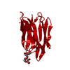
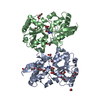
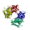
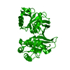
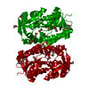
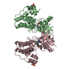
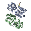
| Title | CRYSTAL STRUCTURE OF HELIANTHUS TUBEROSUS LECTIN COMPLEXED TO MAN(1-2)MAN | |||||||||
 Components Components | AGGLUTININ | |||||||||
 Keywords Keywords | SUGAR BINDING PROTEIN / AGGLUTININ / BETA-PRISM / MANNOSE / JACALIN-RELATED | |||||||||
| Function / homology |  Function and homology information Function and homology information | |||||||||
| Biological species |  | |||||||||
| Method |  X-RAY DIFFRACTION / Resolution: 2.45 Å X-RAY DIFFRACTION / Resolution: 2.45 Å | |||||||||
 Authors Authors | Bourne, Y. / Zamboni, V. / Barre, A. / Peumans, W.J. / van Damme, E.J.M. / Rouge, P. | |||||||||
 Citation Citation | Journal: Structure Fold.Des. / Year: 1999 Title: Helianthus tuberosus lectin reveals a widespread scaffold for mannose-binding lectins. Authors: Bourne, Y. / Zamboni, V. / Barre, A. / Peumans, W.J. / Van Damme, E.J. / Rouge, P. | |||||||||
| History |
|
- Structure visualization
| Structure viewer | Molecule: MolmilJmol/JSmol |
|---|
- Downloads & links
-Download
| PDBx/mmCIF format | 1c3n.cif.gz | 41.8 KB | Display | PDBx/mmCIF format |
|---|---|---|---|---|
| PDB format | pdb1c3n.ent.gz | 29.1 KB | Display | PDB format |
| PDBx/mmJSON format | 1c3n.json.gz | Tree view | PDBx/mmJSON format | |
| Others |  Other downloads Other downloads |
-Validation report
| Arichive directory | https://data.pdbj.org/pub/pdb/validation_reports/c3/1c3nftp://data.pdbj.org/pub/pdb/validation_reports/c3/1c3n | HTTPS FTP |
|---|
-Related structure data


-Links
 PDBj
PDBj

- Assembly
| Deposited unit | 
| ||||||||
|---|---|---|---|---|---|---|---|---|---|
| 1 | 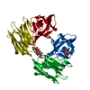
| ||||||||
| 2 | x 8
| ||||||||
| Unit cell |
| ||||||||
| Components on special symmetry positions |
|
-Components
| #1: Protein | Mass: 15501.473 Da / Num. of mol.: 1 / Source method: isolated from a natural source / Details: TUBER Source: (natural) References: GenBank: AAD11575, UniProt: Q9ZQY5*PLUS |
|---|---|
| #2: Polysaccharide | alpha-D-mannopyranose-(1-2)-alpha-D-mannopyranose / 2alpha-alpha-mannobiose  Source method: isolated from a genetically manipulated source Details: oligosaccharide / References: 2alpha-alpha-mannobiose |
| #3: Water | ChemComp-HOH /  Mass: 18.015 Da / Num. of mol.: 88 / Source method: isolated from a natural source / Formula: H2O Mass: 18.015 Da / Num. of mol.: 88 / Source method: isolated from a natural source / Formula: H2O |
-Experimental details
-Experiment
| Experiment | Method: X-RAY DIFFRACTION / Number of used crystals: 1 |
|---|
- Sample preparation
| Crystal | Density Matthews: 2.85 Å3/Da / Density % sol: 56.82 % | |||||||||||||||||||||||||||||||||||||||||||||||||||||||||||||||
|---|---|---|---|---|---|---|---|---|---|---|---|---|---|---|---|---|---|---|---|---|---|---|---|---|---|---|---|---|---|---|---|---|---|---|---|---|---|---|---|---|---|---|---|---|---|---|---|---|---|---|---|---|---|---|---|---|---|---|---|---|---|---|---|---|
| Crystal grow | Temperature: 298 K / Method: vapor diffusion / pH: 7 Details: 26-28% PEG MONOMETHYL ESTER 0.2 M MAGNESIUM ACETATE, pH 7.0, VAPOR DIFFUSION, temperature 298K | |||||||||||||||||||||||||||||||||||||||||||||||||||||||||||||||
| Crystal grow | *PLUS Temperature: 20 ℃ | |||||||||||||||||||||||||||||||||||||||||||||||||||||||||||||||
| Components of the solutions | *PLUS
|
-Data collection
| Diffraction | Mean temperature: 100 K |
|---|---|
| Diffraction source | Source: ROTATING ANODE / Type: RIGAKU RU200 / Wavelength: 1.5418 |
| Detector | Type: MARRESEARCH / Detector: IMAGE PLATE / Date: Jul 14, 1999 |
| Radiation | Protocol: SINGLE WAVELENGTH / Monochromatic (M) / Laue (L): M / Scattering type: x-ray |
| Radiation wavelength | Wavelength: 1.5418 Å / Relative weight: 1 |
| Reflection | Resolution: 2.45→12 Å / Num. obs: 6269 / % possible obs: 91.8 % / Observed criterion σ(F): 0 / Observed criterion σ(I): 0 / Redundancy: 2.4 % / Biso Wilson estimate: 42.4 Å2 / Rmerge(I) obs: 0.083 / Net I/σ(I): 7.1 |
| Reflection shell | Resolution: 2.45→2.55 Å / Redundancy: 2.2 % / Rmerge(I) obs: 0.35 / % possible all: 93.3 |
| Reflection | *PLUS Num. measured all: 53576 |
| Reflection shell | *PLUS % possible obs: 91.7 % / Mean I/σ(I) obs: 2 |
- Processing
| Software |
| ||||||||||||||||||||||||||||||||||||||||||||||||||||||||||||
|---|---|---|---|---|---|---|---|---|---|---|---|---|---|---|---|---|---|---|---|---|---|---|---|---|---|---|---|---|---|---|---|---|---|---|---|---|---|---|---|---|---|---|---|---|---|---|---|---|---|---|---|---|---|---|---|---|---|---|---|---|---|
| Refinement | Resolution: 2.45→12 Å / σ(F): 0 / σ(I): 0 / Stereochemistry target values: ENGH & HUBER
| ||||||||||||||||||||||||||||||||||||||||||||||||||||||||||||
| Refinement step | Cycle: LAST / Resolution: 2.45→12 Å
| ||||||||||||||||||||||||||||||||||||||||||||||||||||||||||||
| Refine LS restraints |
| ||||||||||||||||||||||||||||||||||||||||||||||||||||||||||||
| Software | *PLUS Name: 'CNS' / Classification: refinement | ||||||||||||||||||||||||||||||||||||||||||||||||||||||||||||
| Refinement | *PLUS Num. reflection obs: 6269 | ||||||||||||||||||||||||||||||||||||||||||||||||||||||||||||
| Solvent computation | *PLUS | ||||||||||||||||||||||||||||||||||||||||||||||||||||||||||||
| Displacement parameters | *PLUS | ||||||||||||||||||||||||||||||||||||||||||||||||||||||||||||
| Refine LS restraints | *PLUS
|
