 Movie
Movie Controller

[English] 日本語
 Yorodumi
- PDB-1c39: STRUCTURE OF CATION-DEPENDENT MANNOSE 6-PHOSPHATE RECEPTOR BOUND ... -
+ Open data

- Basic information
| Entry | Database: PDB / ID: 1c39 | |||||||||
|---|---|---|---|---|---|---|---|---|---|---|
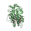
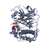
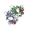
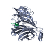
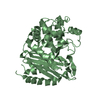
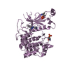
| Title | STRUCTURE OF CATION-DEPENDENT MANNOSE 6-PHOSPHATE RECEPTOR BOUND TO PENTAMANNOSYL PHOSPHATE | |||||||||
 Components Components | CATION-DEPENDENT MANNOSE-6-PHOSPHATE RECEPTOR | |||||||||
 Keywords Keywords | SIGNALING PROTEIN / RECEPTOR / CATION DEPENDENT MANNOSE 6-PHOSPHATE / P-TYPE LECT TRANSPORT | |||||||||
| Function / homology |  Function and homology information Function and homology informationRetrograde transport at the Trans-Golgi-Network / Glycosphingolipid catabolism / secretion of lysosomal enzymes / Lysosome Vesicle Biogenesis / retromer complex binding / Cargo recognition for clathrin-mediated endocytosis / Clathrin-mediated endocytosis / protein targeting to lysosome / lysosomal transport / trans-Golgi network ...Retrograde transport at the Trans-Golgi-Network / Glycosphingolipid catabolism / secretion of lysosomal enzymes / Lysosome Vesicle Biogenesis / retromer complex binding / Cargo recognition for clathrin-mediated endocytosis / Clathrin-mediated endocytosis / protein targeting to lysosome / lysosomal transport / trans-Golgi network / late endosome / endosome / protein domain specific binding / lysosomal membrane / perinuclear region of cytoplasm / Golgi apparatus Similarity search - Function | |||||||||
| Biological species |  | |||||||||
| Method |  X-RAY DIFFRACTION / MOLECULAR REPLACEMEN / Resolution: 1.85 Å X-RAY DIFFRACTION / MOLECULAR REPLACEMEN / Resolution: 1.85 Å | |||||||||
 Authors Authors | Olson, L.J. / Zhang, J. / Lee, Y.C. / Dahms, N.M. / Kim, J.J.-P. | |||||||||
 Citation Citation | Journal: J.Biol.Chem. / Year: 1999 Title: Structural basis for recognition of phosphorylated high mannose oligosaccharides by the cation-dependent mannose 6-phosphate receptor. Authors: Olson, L.J. / Zhang, J. / Lee, Y.C. / Dahms, N.M. / Kim, J.J. #1: Journal: Cell(Cambridge,Mass.) / Year: 1998Title: Molecular Basis of Lysosomal Enzyme Recognition:Enzyme Recognition: Three- Dimensional Structure of the Cation-Dependent Mannose 6-Phosphate Receptor Authors: Roberts, D.L. / Weix, D.J. / Dahms, N.M. / Kim, J.J. | |||||||||
| History |
|
- Structure visualization
| Structure viewer | Molecule: MolmilJmol/JSmol |
|---|
- Downloads & links
-Download
| PDBx/mmCIF format | 1c39.cif.gz | 81.7 KB | Display | PDBx/mmCIF format |
|---|---|---|---|---|
| PDB format | pdb1c39.ent.gz | 60.5 KB | Display | PDB format |
| PDBx/mmJSON format | 1c39.json.gz | Tree view | PDBx/mmJSON format | |
| Others |  Other downloads Other downloads |
-Validation report
| Arichive directory | https://data.pdbj.org/pub/pdb/validation_reports/c3/1c39ftp://data.pdbj.org/pub/pdb/validation_reports/c3/1c39 | HTTPS FTP |
|---|
-Related structure data
| Related structure data | 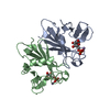 1m6pS S: Starting model for refinement |
|---|---|
| Similar structure data |


-Links
 PDBj
PDBj- Assembly
| Deposited unit | 
| ||||||||
|---|---|---|---|---|---|---|---|---|---|
| 1 |
| ||||||||
| Unit cell |
|
-Components
| #1: Protein | Mass: 17213.363 Da / Num. of mol.: 2 / Fragment: EXTRACYTOPLASMIC DOMAIN / Mutation: N31Q, N57Q, N68Q, N87Q Source method: isolated from a genetically manipulated source Source: (gene. exp.)  Trichoplusia ni (cabbage looper) / References: UniProt: P11456 Trichoplusia ni (cabbage looper) / References: UniProt: P11456#2: Polysaccharide | Source method: isolated from a genetically manipulated source #3: Polysaccharide | Source method: isolated from a genetically manipulated source #4: Chemical |   Mass: 54.938 Da / Num. of mol.: 2 / Source method: obtained synthetically / Formula: Mn Mass: 54.938 Da / Num. of mol.: 2 / Source method: obtained synthetically / Formula: Mn#5: Water | ChemComp-HOH / |  Mass: 18.015 Da / Num. of mol.: 171 / Source method: isolated from a natural source / Formula: H2O Mass: 18.015 Da / Num. of mol.: 171 / Source method: isolated from a natural source / Formula: H2OHas protein modification | Y | |
|---|
-Experimental details
-Experiment
| Experiment | Method: X-RAY DIFFRACTION / Number of used crystals: 1 |
|---|
- Sample preparation
| Crystal | Density Matthews: 2.7 Å3/Da / Density % sol: 54.4 % | ||||||||||||||||||||||||||||||
|---|---|---|---|---|---|---|---|---|---|---|---|---|---|---|---|---|---|---|---|---|---|---|---|---|---|---|---|---|---|---|---|
| Crystal grow | pH: 6.4 / Details: pH 6.40 | ||||||||||||||||||||||||||||||
| Crystal grow | *PLUS Temperature: 19 ℃ / pH: 6.5 / Method: vapor diffusion, hanging drop | ||||||||||||||||||||||||||||||
| Components of the solutions | *PLUS
|
-Data collection
| Diffraction | Mean temperature: 277 K |
|---|---|
| Diffraction source | Source: ROTATING ANODE / Type: RIGAKU RU200 / Wavelength: 1.5418 |
| Detector | Type: RIGAKU RAXIS II / Detector: IMAGE PLATE / Date: Jan 10, 1998 |
| Radiation | Protocol: SINGLE WAVELENGTH / Monochromatic (M) / Laue (L): M / Scattering type: x-ray |
| Radiation wavelength | Wavelength: 1.5418 Å / Relative weight: 1 |
| Reflection | Resolution: 1.85→40 Å / Num. obs: 48273 / % possible obs: 88.4 % / Observed criterion σ(I): 0 / Redundancy: 1.7 % / Biso Wilson estimate: 17.1 Å2 / Rmerge(I) obs: 0.051 / Net I/σ(I): 13.5 |
| Reflection shell | Resolution: 1.85→1.9 Å / Redundancy: 1.3 % / Rmerge(I) obs: 0.292 / % possible all: 62.3 |
| Reflection | *PLUS Num. obs: 28822 / Num. measured all: 48273 |
| Reflection shell | *PLUS % possible obs: 62.3 % |
- Processing
| Software |
| ||||||||||||||||||||||||||||||||||||||||||||||||||||||||||||||||||||||||||||||||
|---|---|---|---|---|---|---|---|---|---|---|---|---|---|---|---|---|---|---|---|---|---|---|---|---|---|---|---|---|---|---|---|---|---|---|---|---|---|---|---|---|---|---|---|---|---|---|---|---|---|---|---|---|---|---|---|---|---|---|---|---|---|---|---|---|---|---|---|---|---|---|---|---|---|---|---|---|---|---|---|---|---|
| Refinement | Method to determine structure: MOLECULAR REPLACEMEN Starting model: PDB ENTRY 1M6P Resolution: 1.85→40 Å / Rfactor Rfree error: 0.005 / Data cutoff high absF: 146959.16 / Data cutoff low absF: 0 / Isotropic thermal model: RESTRAINED / Cross valid method: THROUGHOUT / σ(F): 2 / Details: SIMULATED ANNEALING
| ||||||||||||||||||||||||||||||||||||||||||||||||||||||||||||||||||||||||||||||||
| Displacement parameters | Biso mean: 29.8 Å2
| ||||||||||||||||||||||||||||||||||||||||||||||||||||||||||||||||||||||||||||||||
| Refine analyze |
| ||||||||||||||||||||||||||||||||||||||||||||||||||||||||||||||||||||||||||||||||
| Refinement step | Cycle: LAST / Resolution: 1.85→40 Å
| ||||||||||||||||||||||||||||||||||||||||||||||||||||||||||||||||||||||||||||||||
| Refine LS restraints |
| ||||||||||||||||||||||||||||||||||||||||||||||||||||||||||||||||||||||||||||||||
| LS refinement shell | Resolution: 1.85→1.97 Å / Rfactor Rfree error: 0.02 / Total num. of bins used: 6
| ||||||||||||||||||||||||||||||||||||||||||||||||||||||||||||||||||||||||||||||||
| Xplor file |
| ||||||||||||||||||||||||||||||||||||||||||||||||||||||||||||||||||||||||||||||||
| Software | *PLUS Name: X-PLOR / Version: 3.851 / Classification: refinement | ||||||||||||||||||||||||||||||||||||||||||||||||||||||||||||||||||||||||||||||||
| Refine LS restraints | *PLUS
|
