 Movie
Movie Controller


+ Open data

- Basic information
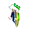
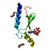
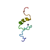
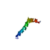
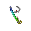
| Entry | Database: PDB / ID: 1bx7 | ||||||
|---|---|---|---|---|---|---|---|
| Title | HIRUSTASIN FROM HIRUDO MEDICINALIS AT 1.2 ANGSTROMS | ||||||
 Components Components | HIRUSTASIN | ||||||
 Keywords Keywords | ANTI-COAGULANT / PEPTIDIC INHIBITORS / CONFORMATIONAL FLEXIBILITY / SERINE PROTEASE INHIBITOR | ||||||
| Function / homology |  Function and homology information Function and homology informationnegative regulation of coagulation / serine-type endopeptidase inhibitor activity / heparin binding / extracellular region Similarity search - Function | ||||||
| Biological species |  Hirudo medicinalis (medicinal leech) Hirudo medicinalis (medicinal leech) | ||||||
| Method |  X-RAY DIFFRACTION / SYNCHROTRON / AB INITIO / Resolution: 1.2 Å X-RAY DIFFRACTION / SYNCHROTRON / AB INITIO / Resolution: 1.2 Å | ||||||
 Authors Authors | Uson, I. / Sheldrick, G.M. / De La Fortelle, E. / Bricogne, G. / Di Marco, S. / Priestle, J.P. / Gruetter, M.G. / Mittl, P.R.E. | ||||||
 Citation Citation | Journal: Structure Fold.Des. / Year: 1999 Title: The 1.2 A crystal structure of hirustasin reveals the intrinsic flexibility of a family of highly disulphide-bridged inhibitors. Authors: Uson, I. / Sheldrick, G.M. / de La Fortelle, E. / Bricogne, G. / Di Marco, S. / Priestle, J.P. / Grutter, M.G. / Mittl, P.R. #1: Journal: Structure / Year: 1997Title: A New Structural Class of Serine Protease Inhibitors Revealed by the Structure of the Hirustasin-Kallikrein Complex Authors: Mittl, P.R. / Di Marco, S. / Fendrich, G. / Pohlig, G. / Heim, J. / Sommerhoff, C. / Fritz, H. / Priestle, J.P. / Grutter, M.G. #2: Journal: Structure / Year: 1997Title: Erratum. A New Structural Class of Serine Protease Inhibitors Revealed by the Structure of the Hirustasin-Kallikrein Complex Authors: Mittl, P.R. / Di Marco, S. / Fendrich, G. / Pohlig, G. / Heim, J. / Sommerhoff, C. / Fritz, H. / Priestle, J.P. / Grutter, M.G. #3: Journal: Protein Sci. / Year: 1997Title: Recombinant Hirustasin: Production in Yeast, Crystallization, and Interaction with Serine Proteases Authors: Di Marco, S. / Fendrich, G. / Knecht, R. / Strauss, A. / Pohlig, G. / Heim, J. / Priestle, J.P. / Sommerhoff, C.P. / Grutter, M.G. | ||||||
| History |
|
- Structure visualization
| Structure viewer | Molecule: MolmilJmol/JSmol |
|---|
- Downloads & links
-Download
| PDBx/mmCIF format | 1bx7.cif.gz | 35 KB | Display | PDBx/mmCIF format |
|---|---|---|---|---|
| PDB format | pdb1bx7.ent.gz | 23.8 KB | Display | PDB format |
| PDBx/mmJSON format | 1bx7.json.gz | Tree view | PDBx/mmJSON format | |
| Others |  Other downloads Other downloads |
-Validation report
| Arichive directory | https://data.pdbj.org/pub/pdb/validation_reports/bx/1bx7ftp://data.pdbj.org/pub/pdb/validation_reports/bx/1bx7 | HTTPS FTP |
|---|
-Related structure data


-Links
 PDBj
PDBj
- Assembly
| Deposited unit | 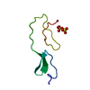
| ||||||||
|---|---|---|---|---|---|---|---|---|---|
| 1 |
| ||||||||
| Unit cell |
| ||||||||
| Components on special symmetry positions |
|
-Components
| #1: Protein | Mass: 5886.788 Da / Num. of mol.: 1 Source method: isolated from a genetically manipulated source Source: (gene. exp.) Hirudo medicinalis (medicinal leech) / Production host:  | ||||
|---|---|---|---|---|---|
| #2: Chemical |   Mass: 96.063 Da / Num. of mol.: 2 / Source method: obtained synthetically / Formula: SO4 Mass: 96.063 Da / Num. of mol.: 2 / Source method: obtained synthetically / Formula: SO4#3: Water | ChemComp-HOH / |  Mass: 18.015 Da / Num. of mol.: 52 / Source method: isolated from a natural source / Formula: H2O Mass: 18.015 Da / Num. of mol.: 52 / Source method: isolated from a natural source / Formula: H2OHas protein modification | Y | |
-Experimental details
-Experiment
| Experiment | Method: X-RAY DIFFRACTION / Number of used crystals: 1 |
|---|
- Sample preparation
| Crystal | Density Matthews: 2 Å3/Da / Density % sol: 40 % Description: TWO DIFFERENT INDEPENDENT METHODS WERE USED FOR STRUCTURE SOLUTION | ||||||||||||||||||||||||
|---|---|---|---|---|---|---|---|---|---|---|---|---|---|---|---|---|---|---|---|---|---|---|---|---|---|
| Crystal grow | pH: 5.45 / Details: pH 5.45 | ||||||||||||||||||||||||
| Crystal | *PLUS | ||||||||||||||||||||||||
| Crystal grow | *PLUS Method: unknown / Details: Di Marco, S., (1997) Protein Sci., 6, 109. | ||||||||||||||||||||||||
| Components of the solutions | *PLUS
|
-Data collection
| Diffraction | Mean temperature: 100 K |
|---|---|
| Diffraction source | Source: SYNCHROTRON / Site: EMBL/DESY, HAMBURG  / Beamline: BW7B / Wavelength: 1.05 / Beamline: BW7B / Wavelength: 1.05 |
| Detector | Type: MARRESEARCH / Detector: IMAGE PLATE / Date: Nov 1, 1996 / Details: BENT CRYSTAL |
| Radiation | Monochromator: GE(111) / Monochromatic (M) / Laue (L): M / Scattering type: x-ray |
| Radiation wavelength | Wavelength: 1.05 Å / Relative weight: 1 |
| Reflection | Resolution: 1.2→25.2 Å / Num. obs: 15928 / % possible obs: 99.4 % / Observed criterion σ(I): 0 / Redundancy: 2.4 % / Rmerge(I) obs: 0.043 / Net I/σ(I): 15 |
| Reflection shell | Resolution: 1.2→1.25 Å / Redundancy: 1.8 % / Rmerge(I) obs: 0.376 / Mean I/σ(I) obs: 1.61 / % possible all: 98.1 |
| Reflection shell | *PLUS % possible obs: 98.1 % |
- Processing
| Software |
| |||||||||||||||||||||||||||||||||
|---|---|---|---|---|---|---|---|---|---|---|---|---|---|---|---|---|---|---|---|---|---|---|---|---|---|---|---|---|---|---|---|---|---|---|
| Refinement | Method to determine structure: AB INITIO / Resolution: 1.2→25.2 Å / Num. parameters: 4045 / Num. restraintsaints: 4895 / Cross valid method: FREE R / σ(F): 0 / Stereochemistry target values: ENGH AND HUBER Details: ANISOTROPIC REFINEMENT REDUCED FREE R (NO CUTOFF) BY 2%
| |||||||||||||||||||||||||||||||||
| Solvent computation | Solvent model: MOEWS & KRETSINGER, J.MOL.BIOL.91(1973)201-2 | |||||||||||||||||||||||||||||||||
| Refine analyze | Num. disordered residues: 5 / Occupancy sum hydrogen: 342 / Occupancy sum non hydrogen: 417.02 | |||||||||||||||||||||||||||||||||
| Refinement step | Cycle: LAST / Resolution: 1.2→25.2 Å
| |||||||||||||||||||||||||||||||||
| Refine LS restraints |
| |||||||||||||||||||||||||||||||||
| Software | *PLUS Name: SHELXL-97 / Classification: refinement | |||||||||||||||||||||||||||||||||
| Refinement | *PLUS Rfactor obs: 0.127 / Rfactor Rwork: 0.1794 | |||||||||||||||||||||||||||||||||
| Solvent computation | *PLUS | |||||||||||||||||||||||||||||||||
| Displacement parameters | *PLUS | |||||||||||||||||||||||||||||||||
| Refine LS restraints | *PLUS
|
