 Movie
Movie Controller


+ Open data

- Basic information
| Entry | Database: PDB / ID: 1bsl | ||||||
|---|---|---|---|---|---|---|---|
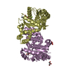
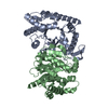
| Title | STRUCTURE OF ALKANAL MONOOXYGENASE BETA CHAIN | ||||||
 Components Components | BACTERIAL LUCIFERASE | ||||||
 Keywords Keywords | FLAVOPROTEIN / PHOTOPROTEIN / LUMINESCENCE / OXIDOREDUCTASE / MONOOXYGENASE / FMN | ||||||
| Function / homology |  Function and homology information Function and homology informationbacterial luciferase / alkanal monooxygenase (FMN-linked) activity / bioluminescence / cytosol Similarity search - Function | ||||||
| Biological species |  Vibrio harveyi (bacteria) Vibrio harveyi (bacteria) | ||||||
| Method |  X-RAY DIFFRACTION / Resolution: 1.95 Å X-RAY DIFFRACTION / Resolution: 1.95 Å | ||||||
 Authors Authors | Rayment, I. / Holden, H.M. / Thoden, J.B. / Baldwin, T.O. | ||||||
 Citation Citation | Journal: Protein Sci. / Year: 1997 Title: Structure of the beta 2 homodimer of bacterial luciferase from Vibrio harveyi: X-ray analysis of a kinetic protein folding trap. Authors: Thoden, J.B. / Holden, H.M. / Fisher, A.J. / Sinclair, J.F. / Wesenberg, G. / Baldwin, T.O. / Rayment, I. #1: Journal: J.Biol.Chem. / Year: 1996Title: The 1.5-A Resolution Crystal Structure of Bacterial Luciferase in Low Salt Conditions Authors: Fisher, A.J. / Thompson, T.B. / Thoden, J.B. / Baldwin, T.O. / Rayment, I. | ||||||
| History |
|
- Structure visualization
| Structure viewer | Molecule: MolmilJmol/JSmol |
|---|
- Downloads & links
-Download
| PDBx/mmCIF format | 1bsl.cif.gz | 160.9 KB | Display | PDBx/mmCIF format |
|---|---|---|---|---|
| PDB format | pdb1bsl.ent.gz | 126.5 KB | Display | PDB format |
| PDBx/mmJSON format | 1bsl.json.gz | Tree view | PDBx/mmJSON format | |
| Others |  Other downloads Other downloads |
-Validation report
| Arichive directory | https://data.pdbj.org/pub/pdb/validation_reports/bs/1bslftp://data.pdbj.org/pub/pdb/validation_reports/bs/1bsl | HTTPS FTP |
|---|
-Related structure data
| Similar structure data |
|---|


-Links
 PDBj
PDBj
- Assembly
| Deposited unit | 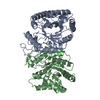
| ||||||||
|---|---|---|---|---|---|---|---|---|---|
| 1 |
| ||||||||
| Unit cell |
|
-Components
| #1: Protein | Mass: 36384.684 Da / Num. of mol.: 2 / Fragment: BETA SUBUNIT Source method: isolated from a genetically manipulated source Source: (gene. exp.) Vibrio harveyi (bacteria) / Production host: #2: Water | ChemComp-HOH / |  Mass: 18.015 Da / Num. of mol.: 989 / Source method: isolated from a natural source / Formula: H2O Mass: 18.015 Da / Num. of mol.: 989 / Source method: isolated from a natural source / Formula: H2O |
|---|
-Experimental details
-Experiment
| Experiment | Method: X-RAY DIFFRACTION |
|---|
- Sample preparation
| Crystal | Density Matthews: 2.73 Å3/Da / Density % sol: 54.98 % | |||||||||||||||||||||||||||||||||||||||||||||
|---|---|---|---|---|---|---|---|---|---|---|---|---|---|---|---|---|---|---|---|---|---|---|---|---|---|---|---|---|---|---|---|---|---|---|---|---|---|---|---|---|---|---|---|---|---|---|
| Crystal grow | *PLUS pH: 7 / Method: vapor diffusion, hanging drop | |||||||||||||||||||||||||||||||||||||||||||||
| Components of the solutions | *PLUS
|
-Data collection
| Diffraction source | Wavelength: 1.5418 |
|---|---|
| Detector | Type: SIEMENS / Detector: AREA DETECTOR / Date: Jun 1, 1995 |
| Radiation | Monochromatic (M) / Laue (L): M / Scattering type: x-ray |
| Radiation wavelength | Wavelength: 1.5418 Å / Relative weight: 1 |
| Reflection | Num. obs: 55442 / % possible obs: 93.7 % / Redundancy: 2.2 % / Rmerge(I) obs: 0.041 |
| Reflection | *PLUS Highest resolution: 1.95 Å / Lowest resolution: 30 Å / Num. measured all: 125700 |
| Reflection shell | *PLUS Highest resolution: 1.95 Å / % possible obs: 85.1 % / Num. unique obs: 6873 / Num. measured obs: 9563 / Rmerge(I) obs: 0.18 |
- Processing
| Software |
| ||||||||||||||||||||||||||||||
|---|---|---|---|---|---|---|---|---|---|---|---|---|---|---|---|---|---|---|---|---|---|---|---|---|---|---|---|---|---|---|---|
| Refinement | Resolution: 1.95→30 Å /
| ||||||||||||||||||||||||||||||
| Refinement step | Cycle: LAST / Resolution: 1.95→30 Å
| ||||||||||||||||||||||||||||||
| Refine LS restraints |
| ||||||||||||||||||||||||||||||
| Software | *PLUS Name: TNT / Classification: refinement | ||||||||||||||||||||||||||||||
| Refinement | *PLUS Rfactor all: 0.188 | ||||||||||||||||||||||||||||||
| Solvent computation | *PLUS | ||||||||||||||||||||||||||||||
| Displacement parameters | *PLUS | ||||||||||||||||||||||||||||||
| Refine LS restraints | *PLUS
|
