 Movie
Movie Controller

[English] 日本語
 Yorodumi
- PDB-1bq7: DSBA MUTANT P151A, ROLE OF THE CIS-PROLINE IN THE ACTIVE SITE OF DSBA -
+ Open data

- Basic information
| Entry | Database: PDB / ID: 1bq7 | ||||||
|---|---|---|---|---|---|---|---|
| Title | DSBA MUTANT P151A, ROLE OF THE CIS-PROLINE IN THE ACTIVE SITE OF DSBA | ||||||
 Components Components | PROTEIN (DISULFIDE OXIDOREDUCTASE) | ||||||
 Keywords Keywords | OXIDOREDUCTASE / DISULFIDE OXIDOREDUCTASE / PROTEIN DISULFIDE ISOMERASE / PROTEIN FOLDING / REDOX PROTEIN / REDOX-ACTIVE CENTER | ||||||
| Function / homology |  Function and homology information Function and homology informationcellular response to antibiotic / protein disulfide isomerase activity / Secretion of toxins / protein-disulfide reductase activity / outer membrane-bounded periplasmic space / periplasmic space / oxidoreductase activity Similarity search - Function | ||||||
| Biological species |  | ||||||
| Method |  X-RAY DIFFRACTION / SYNCHROTRON / MOLECULAR REPLACEMENT / Resolution: 2.8 Å X-RAY DIFFRACTION / SYNCHROTRON / MOLECULAR REPLACEMENT / Resolution: 2.8 Å | ||||||
 Authors Authors | Charbonnier, J.-B. / Stura, E.A. | ||||||
 Citation Citation | Journal: Protein Sci. / Year: 1999 Title: On the role of the cis-proline residue in the active site of DsbA. Authors: Charbonnier, J.B. / Belin, P. / Moutiez, M. / Stura, E.A. / Quemeneur, E. #1: Journal: Protein Sci. / Year: 1997Title: Structural Analysis of Three His32 Mutants of DsbA: Support for an Electrostatic Role of His32 in DsbA Stability Authors: Guddat, L. / Bardwell, J.C. / Glockshuber, R. / Huber-Wunderlich, M. / Zander, T. / Martin, J.L. | ||||||
| History |
|
- Structure visualization
| Structure viewer | Molecule: MolmilJmol/JSmol |
|---|
- Downloads & links
-Download
| PDBx/mmCIF format | 1bq7.cif.gz | 198.3 KB | Display | PDBx/mmCIF format |
|---|---|---|---|---|
| PDB format | pdb1bq7.ent.gz | 164.3 KB | Display | PDB format |
| PDBx/mmJSON format | 1bq7.json.gz | Tree view | PDBx/mmJSON format | |
| Others |  Other downloads Other downloads |
-Validation report
| Arichive directory | https://data.pdbj.org/pub/pdb/validation_reports/bq/1bq7ftp://data.pdbj.org/pub/pdb/validation_reports/bq/1bq7 | HTTPS FTP |
|---|
-Related structure data
| Related structure data |  1fvkS S: Starting model for refinement |
|---|---|
| Similar structure data |


-Links
 PDBj
PDBj

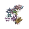
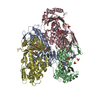
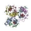
- Assembly
| Deposited unit | 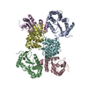
| ||||||||||||||||||||||||||||||||||||
|---|---|---|---|---|---|---|---|---|---|---|---|---|---|---|---|---|---|---|---|---|---|---|---|---|---|---|---|---|---|---|---|---|---|---|---|---|---|
| 1 |
| ||||||||||||||||||||||||||||||||||||
| Unit cell |
| ||||||||||||||||||||||||||||||||||||
| Noncrystallographic symmetry (NCS) | NCS domain:
NCS oper:
|
-Components
| #1: Protein | Mass: 21128.988 Da / Num. of mol.: 6 / Mutation: P151A Source method: isolated from a genetically manipulated source Source: (gene. exp.) Compound details | THERE ARE SIX MOLECULES IN THE ASYMMETRIC UNIT. EACH CONTAINS 186 RESIDUES THE ACTIVE SITE ...THERE ARE SIX MOLECULES IN THE ASYMMETRIC | Has protein modification | Y | |
|---|
-Experimental details
-Experiment
| Experiment | Method: X-RAY DIFFRACTION / Number of used crystals: 1 |
|---|
- Sample preparation
| Crystal | Density Matthews: 3.05 Å3/Da / Density % sol: 59.51 % | ||||||||||||||||||||||||||||||||||||||||
|---|---|---|---|---|---|---|---|---|---|---|---|---|---|---|---|---|---|---|---|---|---|---|---|---|---|---|---|---|---|---|---|---|---|---|---|---|---|---|---|---|---|
| Crystal grow | pH: 6.5 Details: 25% PEG 8,000 100 MM NACL 100 MM SODIUM CACODYLATE, PH 6.5 10% DMSO DIFFUSION VAPOR AT ROOM TEMPERATURE, SEEDING | ||||||||||||||||||||||||||||||||||||||||
| Crystal grow | *PLUS Temperature: 21 ℃ / Method: vapor diffusion, hanging drop | ||||||||||||||||||||||||||||||||||||||||
| Components of the solutions | *PLUS
|
-Data collection
| Diffraction | Mean temperature: 289 K |
|---|---|
| Diffraction source | Source: SYNCHROTRON / Site: LURE  / Beamline: DW32 / Wavelength: 0.97 / Beamline: DW32 / Wavelength: 0.97 |
| Detector | Date: Jul 15, 1997 |
| Radiation | Protocol: SINGLE WAVELENGTH / Monochromatic (M) / Laue (L): M / Scattering type: x-ray |
| Radiation wavelength | Wavelength: 0.97 Å / Relative weight: 1 |
| Reflection | Resolution: 2.8→20 Å / Num. obs: 31364 / % possible obs: 84.6 % / Observed criterion σ(I): 0.3 / Redundancy: 3.8 % / Rmerge(I) obs: 0.078 / Rsym value: 0.078 / Net I/σ(I): 13.3 |
| Reflection shell | Resolution: 2.8→2.9 Å / Rmerge(I) obs: 0.475 / Mean I/σ(I) obs: 2.3 / Rsym value: 0.475 / % possible all: 87.7 |
| Reflection | *PLUS Num. measured all: 117916 |
| Reflection shell | *PLUS % possible obs: 87.9 % / Mean I/σ(I) obs: 1.7 |
- Processing
| Software |
| ||||||||||||||||||||||||||||||||||||||||||||||||||||||||||||
|---|---|---|---|---|---|---|---|---|---|---|---|---|---|---|---|---|---|---|---|---|---|---|---|---|---|---|---|---|---|---|---|---|---|---|---|---|---|---|---|---|---|---|---|---|---|---|---|---|---|---|---|---|---|---|---|---|---|---|---|---|---|
| Refinement | Method to determine structure: MOLECULAR REPLACEMENT Starting model: PDB ENTRY 1FVK Resolution: 2.8→10 Å / Cross valid method: THROUGHOUT / σ(F): 1 Details: PRO 151 IS MUTATED TO ALANINE. PEPTIDE BOND VAL150-PRO151 IS IN CIS CONFORMATION IN WILD TYPE DSBA. IT IS IN TRANS-CONFORMATION IN MUTANT DSBA PRO 151 ALA.
| ||||||||||||||||||||||||||||||||||||||||||||||||||||||||||||
| Displacement parameters | Biso mean: 53.7 Å2 | ||||||||||||||||||||||||||||||||||||||||||||||||||||||||||||
| Refinement step | Cycle: LAST / Resolution: 2.8→10 Å
| ||||||||||||||||||||||||||||||||||||||||||||||||||||||||||||
| Refine LS restraints |
| ||||||||||||||||||||||||||||||||||||||||||||||||||||||||||||
| Refine LS restraints NCS |
| ||||||||||||||||||||||||||||||||||||||||||||||||||||||||||||
| LS refinement shell | Resolution: 2.8→2.92 Å
| ||||||||||||||||||||||||||||||||||||||||||||||||||||||||||||
| Software | *PLUS Name: X-PLOR / Version: 3.1 / Classification: refinement | ||||||||||||||||||||||||||||||||||||||||||||||||||||||||||||
| Refinement | *PLUS Highest resolution: 2.8 Å / σ(F): 1 / % reflection Rfree: 5 % | ||||||||||||||||||||||||||||||||||||||||||||||||||||||||||||
| Solvent computation | *PLUS | ||||||||||||||||||||||||||||||||||||||||||||||||||||||||||||
| Displacement parameters | *PLUS Biso mean: 53.7 Å2 | ||||||||||||||||||||||||||||||||||||||||||||||||||||||||||||
| Refine LS restraints | *PLUS
| ||||||||||||||||||||||||||||||||||||||||||||||||||||||||||||
| LS refinement shell | *PLUS Highest resolution: 2.8 Å / Rfactor Rfree: 0.418 / % reflection Rfree: 4.5 % / Rfactor Rwork: 0.348 |
