 Movie
Movie Controller

[English] 日本語
 Yorodumi
- PDB-1b76: GLYCYL-TRNA SYNTHETASE FROM THERMUS THERMOPHILUS COMPLEXED WITH ATP -
+ Open data

- Basic information
| Entry | Database: PDB / ID: 1b76 | ||||||
|---|---|---|---|---|---|---|---|
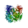
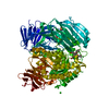
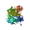
| Title | GLYCYL-TRNA SYNTHETASE FROM THERMUS THERMOPHILUS COMPLEXED WITH ATP | ||||||
 Components Components | Glycine--tRNA ligase | ||||||
 Keywords Keywords | LIGASE / AMINOACYL-TRNA SYNTHASE / LIGASE(SYNTHETASE) | ||||||
| Function / homology |  Function and homology information Function and homology informationglycine-tRNA ligase complex / glycyl-tRNA aminoacylation / glycine-tRNA ligase / glycine-tRNA ligase activity / glycine binding / protein dimerization activity / magnesium ion binding / ATP binding / identical protein binding Similarity search - Function | ||||||
| Biological species |   Thermus thermophilus (bacteria) Thermus thermophilus (bacteria) | ||||||
| Method |  X-RAY DIFFRACTION / SYNCHROTRON / OTHER / Resolution: 3.4 Å X-RAY DIFFRACTION / SYNCHROTRON / OTHER / Resolution: 3.4 Å | ||||||
 Authors Authors | Arnez, J.G. / Moras, D. | ||||||
 Citation Citation | Journal: J.Mol.Biol. / Year: 1999 Title: Glycyl-tRNA synthetase uses a negatively charged pit for specific recognition and activation of glycine. Authors: Arnez, J.G. / Dock-Bregeon, A.C. / Moras, D. #1: Journal: Embo J. / Year: 1995Title: Crystal structure of glycyl-tRNA synthetase from Thermus thermophilus. Authors: Logan, D.T. / Mazauric, M.H. / Kern, D. / Moras, D. #2: Journal: J.Mol.Biol. / Year: 1994 Title: Crystallisation of the glycyl-tRNA synthetase from Thermus thermophilus and initial crystallographic data. Authors: Logan, D.T. / Cura, V. / Touzel, J.P. / Kern, D. / Moras, D. | ||||||
| History |
|
- Structure visualization
| Structure viewer | Molecule: MolmilJmol/JSmol |
|---|
- Downloads & links
-Download
| PDBx/mmCIF format | 1b76.cif.gz | 225.3 KB | Display | PDBx/mmCIF format |
|---|---|---|---|---|
| PDB format | pdb1b76.ent.gz | 180.1 KB | Display | PDB format |
| PDBx/mmJSON format | 1b76.json.gz | Tree view | PDBx/mmJSON format | |
| Others |  Other downloads Other downloads |
-Validation report
| Arichive directory | https://data.pdbj.org/pub/pdb/validation_reports/b7/1b76ftp://data.pdbj.org/pub/pdb/validation_reports/b7/1b76 | HTTPS FTP |
|---|
-Related structure data


-Links
 PDBj
PDBj
- Assembly
| Deposited unit | 
| ||||||||||
|---|---|---|---|---|---|---|---|---|---|---|---|
| 1 |
| ||||||||||
| Unit cell |
| ||||||||||
| Noncrystallographic symmetry (NCS) | NCS oper: (Code: given Matrix: (-0.634886, -0.710321, 0.303914), Vector: |
-Components
| #1: Protein | Mass: 51144.949 Da / Num. of mol.: 2 Source method: isolated from a genetically manipulated source Details: ATP BOUND IN ACTIVE SITE OF EACH MONOMER Source: (gene. exp.) Thermus thermophilus (strain HB8 / ATCC 27634 / DSM 579) (bacteria)Species: Thermus thermophilus / Strain: HB8 / ATCC 27634 / DSM 579 / Gene: glyQS, glyS, TTHA0543 / Plasmid: PGRS / Gene (production host): GLYS / Production host: #2: Chemical |   Mass: 507.181 Da / Num. of mol.: 2 / Source method: obtained synthetically / Formula: C10H16N5O13P3 / Comment: ATP, energy-carrying molecule*YM Mass: 507.181 Da / Num. of mol.: 2 / Source method: obtained synthetically / Formula: C10H16N5O13P3 / Comment: ATP, energy-carrying molecule*YM#3: Water | ChemComp-HOH / |  Mass: 18.015 Da / Num. of mol.: 2 / Source method: isolated from a natural source / Formula: H2O Mass: 18.015 Da / Num. of mol.: 2 / Source method: isolated from a natural source / Formula: H2OSequence details | A P56206 96 - 158 GAP IN PDB ENTRY B P56206 96 - 158 GAP IN PDB ENTRY | |
|---|
-Experimental details
-Experiment
| Experiment | Method: X-RAY DIFFRACTION / Number of used crystals: 5 |
|---|
- Sample preparation
| Crystal | Density Matthews: 3.6 Å3/Da / Density % sol: 61 % | ||||||||||||||||||||||||||||||||||||||||||||||||||||||||
|---|---|---|---|---|---|---|---|---|---|---|---|---|---|---|---|---|---|---|---|---|---|---|---|---|---|---|---|---|---|---|---|---|---|---|---|---|---|---|---|---|---|---|---|---|---|---|---|---|---|---|---|---|---|---|---|---|---|
| Crystal grow | Temperature: 297 K / pH: 8 / Details: pH 8.0, temperature 297K | ||||||||||||||||||||||||||||||||||||||||||||||||||||||||
| Components of the solutions |
| ||||||||||||||||||||||||||||||||||||||||||||||||||||||||
| Crystal grow | *PLUS Temperature: 24 ℃ / Method: vapor diffusion, hanging drop | ||||||||||||||||||||||||||||||||||||||||||||||||||||||||
| Components of the solutions | *PLUS
|
-Data collection
| Diffraction | Mean temperature: 272 K |
|---|---|
| Diffraction source | Source: SYNCHROTRON / Site: LURE  / Beamline: DW32 / Wavelength: 0.98 / Beamline: DW32 / Wavelength: 0.98 |
| Detector | Type: MARRESEARCH / Detector: IMAGE PLATE / Date: Apr 23, 1996 |
| Radiation | Protocol: SINGLE WAVELENGTH / Monochromatic (M) / Laue (L): M / Scattering type: x-ray |
| Radiation wavelength | Wavelength: 0.98 Å / Relative weight: 1 |
| Reflection | Resolution: 3.3→15 Å / Num. all: 20177 / Num. obs: 20177 / % possible obs: 81 % / Observed criterion σ(I): 0 / Redundancy: 3.4 % / Rmerge(I) obs: 0.095 / Rsym value: 9.5 |
| Reflection | *PLUS |
- Processing
| Software |
| ||||||||||||||||||||||||||||||||||||||||||||||||||||||||||||
|---|---|---|---|---|---|---|---|---|---|---|---|---|---|---|---|---|---|---|---|---|---|---|---|---|---|---|---|---|---|---|---|---|---|---|---|---|---|---|---|---|---|---|---|---|---|---|---|---|---|---|---|---|---|---|---|---|---|---|---|---|---|
| Refinement | Method to determine structure: OTHER / Resolution: 3.4→9 Å / Isotropic thermal model: RESTRAINED / Cross valid method: THROUGHOUT / σ(F): 2 / Details: FINAL RMS COORD. SHIFT 0.012 ANGSTROMS
| ||||||||||||||||||||||||||||||||||||||||||||||||||||||||||||
| Displacement parameters | Biso mean: 43.4 Å2 | ||||||||||||||||||||||||||||||||||||||||||||||||||||||||||||
| Refine analyze | Luzzati d res low obs: 9 Å | ||||||||||||||||||||||||||||||||||||||||||||||||||||||||||||
| Refinement step | Cycle: LAST / Resolution: 3.4→9 Å
| ||||||||||||||||||||||||||||||||||||||||||||||||||||||||||||
| Refine LS restraints |
| ||||||||||||||||||||||||||||||||||||||||||||||||||||||||||||
| Refine LS restraints NCS | NCS model details: RESTRAINTS | ||||||||||||||||||||||||||||||||||||||||||||||||||||||||||||
| LS refinement shell | Resolution: 3.4→3.55 Å / Total num. of bins used: 8
| ||||||||||||||||||||||||||||||||||||||||||||||||||||||||||||
| Xplor file |
| ||||||||||||||||||||||||||||||||||||||||||||||||||||||||||||
| Software | *PLUS Name: X-PLOR / Classification: refinement | ||||||||||||||||||||||||||||||||||||||||||||||||||||||||||||
| Refinement | *PLUS Highest resolution: 3.4 Å / Lowest resolution: 9 Å / σ(F): 2 / % reflection Rfree: 4 % / Rfactor obs: 0.227 | ||||||||||||||||||||||||||||||||||||||||||||||||||||||||||||
| Solvent computation | *PLUS | ||||||||||||||||||||||||||||||||||||||||||||||||||||||||||||
| Displacement parameters | *PLUS Biso mean: 43.4 Å2 | ||||||||||||||||||||||||||||||||||||||||||||||||||||||||||||
| Refine LS restraints | *PLUS
| ||||||||||||||||||||||||||||||||||||||||||||||||||||||||||||
| LS refinement shell | *PLUS Highest resolution: 3.4 Å / % reflection Rfree: 4 % |
