 Movie
Movie Controller

[English] 日本語
 Yorodumi
- PDB-1g5h: CRYSTAL STRUCTURE OF THE ACCESSORY SUBUNIT OF MURINE MITOCHONDRIA... -
+ Open data

- Basic information
| Entry | Database: PDB / ID: 1g5h | ||||||
|---|---|---|---|---|---|---|---|
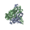
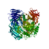
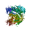
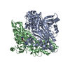
| Title | CRYSTAL STRUCTURE OF THE ACCESSORY SUBUNIT OF MURINE MITOCHONDRIAL POLYMERASE GAMMA | ||||||
 Components Components | MITOCHONDRIAL DNA POLYMERASE ACCESSORY SUBUNIT | ||||||
 Keywords Keywords | DNA BINDING PROTEIN / intermolecular four helix bundle | ||||||
| Function / homology |  Function and homology information Function and homology informationStrand-asynchronous mitochondrial DNA replication / gamma DNA polymerase complex / mitochondrial DNA metabolic process / mitochondrial DNA replication / DNA polymerase processivity factor activity / mitochondrial nucleoid / DNA polymerase binding / mitochondrion organization / DNA-templated DNA replication / double-stranded DNA binding ...Strand-asynchronous mitochondrial DNA replication / gamma DNA polymerase complex / mitochondrial DNA metabolic process / mitochondrial DNA replication / DNA polymerase processivity factor activity / mitochondrial nucleoid / DNA polymerase binding / mitochondrion organization / DNA-templated DNA replication / double-stranded DNA binding / in utero embryonic development / DNA-directed DNA polymerase activity / DNA replication / mitochondrial matrix / DNA repair / mitochondrion / identical protein binding / cytoplasm Similarity search - Function | ||||||
| Biological species |  | ||||||
| Method |  X-RAY DIFFRACTION / SYNCHROTRON / MAD / Resolution: 1.95 Å X-RAY DIFFRACTION / SYNCHROTRON / MAD / Resolution: 1.95 Å | ||||||
 Authors Authors | Carrodeguas, J.A. / Theis, K. / Bogenhagen, D.F. / Kisker, C. | ||||||
 Citation Citation | Journal: Mol.Cell / Year: 2001 Title: Crystal structure and deletion analysis show that the accessory subunit of mammalian DNA polymerase gamma, Pol gamma B, functions as a homodimer. Authors: Carrodeguas, J.A. / Theis, K. / Bogenhagen, D.F. / Kisker, C. #1: Journal: Nucleic Acids Res. / Year: 2000Title: Protein Sequences Conserved in Prokaryotic Aminoacyl-tRNA Synthetases are Important for the Activity of the Processivity Factor of Human Mitochondrial DNA Polymerase Authors: Carrodeguas, J.A. / Bogenhagen, D.F. | ||||||
| History |
|
- Structure visualization
| Structure viewer | Molecule: MolmilJmol/JSmol |
|---|
- Downloads & links
-Download
| PDBx/mmCIF format | 1g5h.cif.gz | 349.1 KB | Display | PDBx/mmCIF format |
|---|---|---|---|---|
| PDB format | pdb1g5h.ent.gz | 283.1 KB | Display | PDB format |
| PDBx/mmJSON format | 1g5h.json.gz | Tree view | PDBx/mmJSON format | |
| Others |  Other downloads Other downloads |
-Validation report
| Arichive directory | https://data.pdbj.org/pub/pdb/validation_reports/g5/1g5hftp://data.pdbj.org/pub/pdb/validation_reports/g5/1g5h | HTTPS FTP |
|---|
-Related structure data


-Links
 PDBj
PDBj
- Assembly
| Deposited unit | 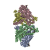
| ||||||||
|---|---|---|---|---|---|---|---|---|---|
| 1 | 
| ||||||||
| 2 | 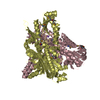
| ||||||||
| Unit cell |
|
-Components
| #1: Protein | Mass: 51336.285 Da / Num. of mol.: 4 Source method: isolated from a genetically manipulated source Source: (gene. exp.)  #2: Chemical | ChemComp-NA /   Mass: 22.990 Da / Num. of mol.: 4 / Source method: obtained synthetically / Formula: Na Mass: 22.990 Da / Num. of mol.: 4 / Source method: obtained synthetically / Formula: Na#3: Chemical |   Mass: 92.094 Da / Num. of mol.: 2 / Source method: obtained synthetically / Formula: C3H8O3 Mass: 92.094 Da / Num. of mol.: 2 / Source method: obtained synthetically / Formula: C3H8O3#4: Water | ChemComp-HOH / |  Mass: 18.015 Da / Num. of mol.: 872 / Source method: isolated from a natural source / Formula: H2O Mass: 18.015 Da / Num. of mol.: 872 / Source method: isolated from a natural source / Formula: H2OHas protein modification | Y | |
|---|
-Experimental details
-Experiment
| Experiment | Method: X-RAY DIFFRACTION / Number of used crystals: 1 |
|---|
- Sample preparation
| Crystal | Density Matthews: 2.12 Å3/Da / Density % sol: 41.94 % | ||||||||||||||||||||||||||||||||||||
|---|---|---|---|---|---|---|---|---|---|---|---|---|---|---|---|---|---|---|---|---|---|---|---|---|---|---|---|---|---|---|---|---|---|---|---|---|---|
| Crystal grow | Temperature: 298 K / Method: vapor diffusion, hanging drop / pH: 7.5 Details: Sodium citrate, pH 7.5, VAPOR DIFFUSION, HANGING DROP, temperature 298K | ||||||||||||||||||||||||||||||||||||
| Crystal grow | *PLUS Details: used microseeding | ||||||||||||||||||||||||||||||||||||
| Components of the solutions | *PLUS
|
-Data collection
| Diffraction |
| |||||||||||||||
|---|---|---|---|---|---|---|---|---|---|---|---|---|---|---|---|---|
| Diffraction source |
| |||||||||||||||
| Detector |
| |||||||||||||||
| Radiation |
| |||||||||||||||
| Radiation wavelength | Wavelength: 1.1 Å / Relative weight: 1 | |||||||||||||||
| Reflection | Resolution: 1.95→20 Å / Num. all: 127289 / Num. obs: 127238 / % possible obs: 99.96 % / Observed criterion σ(F): -3 / Observed criterion σ(I): -3 / Redundancy: 7.4 % / Biso Wilson estimate: 30.741 Å2 / Rmerge(I) obs: 0.072 / Net I/σ(I): 29.6 | |||||||||||||||
| Reflection shell | Resolution: 1.95→2.05 Å / Redundancy: 3.5 % / Rmerge(I) obs: 0.5 / Mean I/σ(I) obs: 2.2 / % possible all: 100 | |||||||||||||||
| Reflection | *PLUS | |||||||||||||||
| Reflection shell | *PLUS % possible obs: 100 % |

- Processing
| Software |
| ||||||||||||||||||||||||||||||||||||||||||||
|---|---|---|---|---|---|---|---|---|---|---|---|---|---|---|---|---|---|---|---|---|---|---|---|---|---|---|---|---|---|---|---|---|---|---|---|---|---|---|---|---|---|---|---|---|---|
| Refinement | Method to determine structure: MAD / Resolution: 1.95→20 Å / SU B: 4.892 / SU ML: 0.144 / Cross valid method: THROUGHOUT / σ(F): 0 / σ(I): -1000 / ESU R: 0.413 / ESU R Free: 0.21 / Stereochemistry target values: standard refmac5
| ||||||||||||||||||||||||||||||||||||||||||||
| Displacement parameters | Biso mean: 42.48 Å2
| ||||||||||||||||||||||||||||||||||||||||||||
| Refinement step | Cycle: LAST / Resolution: 1.95→20 Å
| ||||||||||||||||||||||||||||||||||||||||||||
| Refine LS restraints |
| ||||||||||||||||||||||||||||||||||||||||||||
| Software | *PLUS Name: REFMAC5 / Classification: refinement | ||||||||||||||||||||||||||||||||||||||||||||
| Refinement | *PLUS σ(F): 0 / % reflection Rfree: 2 % | ||||||||||||||||||||||||||||||||||||||||||||
| Solvent computation | *PLUS | ||||||||||||||||||||||||||||||||||||||||||||
| Displacement parameters | *PLUS |
