 Movie
Movie Controller


+ Open data

- Basic information
| Entry | Database: PDB / ID: 1az2 | ||||||
|---|---|---|---|---|---|---|---|
| Title | CITRATE BOUND, C298A/W219Y MUTANT HUMAN ALDOSE REDUCTASE | ||||||
 Components Components | ALDOSE REDUCTASE | ||||||
 Keywords Keywords | OXIDOREDUCTASE / ALDO-KETO REDUCTASE / INHIBITOR BINDING | ||||||
| Function / homology |  Function and homology information Function and homology informationglyceraldehyde oxidoreductase activity / Fructose biosynthesis / fructose biosynthetic process / L-glucuronate reductase activity / aldose reductase / D/L-glyceraldehyde reductase / glycerol dehydrogenase (NADP+) activity / C21-steroid hormone biosynthetic process / NADP-retinol dehydrogenase / Pregnenolone biosynthesis ...glyceraldehyde oxidoreductase activity / Fructose biosynthesis / fructose biosynthetic process / L-glucuronate reductase activity / aldose reductase / D/L-glyceraldehyde reductase / glycerol dehydrogenase (NADP+) activity / C21-steroid hormone biosynthetic process / NADP-retinol dehydrogenase / Pregnenolone biosynthesis / allyl-alcohol dehydrogenase / allyl-alcohol dehydrogenase activity / Galactose catabolism / regulation of urine volume / prostaglandin H2 endoperoxidase reductase activity / all-trans-retinol dehydrogenase (NADP+) activity / metanephric collecting duct development / daunorubicin metabolic process / doxorubicin metabolic process / retinal dehydrogenase (NAD+) activity / epithelial cell maturation / aldose reductase (NADPH) activity / cellular hyperosmotic salinity response / retinoid metabolic process / renal water homeostasis / carbohydrate metabolic process / electron transfer activity / negative regulation of apoptotic process / mitochondrion / : / extracellular exosome / nucleoplasm / cytosol Similarity search - Function | ||||||
| Biological species |  Homo sapiens (human) Homo sapiens (human) | ||||||
| Method |  X-RAY DIFFRACTION / DIFFERENCE FOURIER / Resolution: 2.9 Å X-RAY DIFFRACTION / DIFFERENCE FOURIER / Resolution: 2.9 Å | ||||||
 Authors Authors | Harrison, D.H. / Bohren, K.M. / Ringe, D. / Petsko, G.A. / Gabbay, K.H. | ||||||
 Citation Citation | Journal: Biochemistry / Year: 1997 Title: The alrestatin double-decker: binding of two inhibitor molecules to human aldose reductase reveals a new specificity determinant. Authors: Harrison, D.H. / Bohren, K.M. / Petsko, G.A. / Ringe, D. / Gabbay, K.H. | ||||||
| History |
|
- Structure visualization
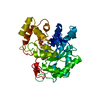
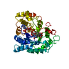
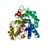
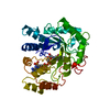
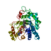
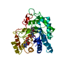
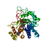
| Structure viewer | Molecule: MolmilJmol/JSmol |
|---|
- Downloads & links
-Download
| PDBx/mmCIF format | 1az2.cif.gz | 68.8 KB | Display | PDBx/mmCIF format |
|---|---|---|---|---|
| PDB format | pdb1az2.ent.gz | 50.1 KB | Display | PDB format |
| PDBx/mmJSON format | 1az2.json.gz | Tree view | PDBx/mmJSON format | |
| Others |  Other downloads Other downloads |
-Validation report
| Arichive directory | https://data.pdbj.org/pub/pdb/validation_reports/az/1az2ftp://data.pdbj.org/pub/pdb/validation_reports/az/1az2 | HTTPS FTP |
|---|
-Related structure data
| Related structure data |  1az1C  2acsS S: Starting model for refinement C: citing same article ( |
|---|---|
| Similar structure data |


-Links
 PDBj
PDBj
- Assembly
| Deposited unit | 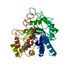
| ||||||||
|---|---|---|---|---|---|---|---|---|---|
| 1 |
| ||||||||
| Unit cell |
|
-Components
| #1: Protein | Mass: 35712.047 Da / Num. of mol.: 1 / Mutation: W219Y, C298A Source method: isolated from a genetically manipulated source Details: CITRATE BOUND / Source: (gene. exp.) Homo sapiens (human) / Cell line: BL21 / Gene: ALR2 / Plasmid: PET / Species (production host): Escherichia coli / Gene (production host): ALR2 / Production host:  |
|---|---|
| #2: Chemical | ChemComp-NAP /   Mass: 743.405 Da / Num. of mol.: 1 / Source method: obtained synthetically / Formula: C21H28N7O17P3 Mass: 743.405 Da / Num. of mol.: 1 / Source method: obtained synthetically / Formula: C21H28N7O17P3 |
| #3: Chemical | ChemComp-CIT /   Mass: 192.124 Da / Num. of mol.: 1 / Source method: obtained synthetically / Formula: C6H8O7 Mass: 192.124 Da / Num. of mol.: 1 / Source method: obtained synthetically / Formula: C6H8O7 |
-Experimental details
-Experiment
| Experiment | Method: X-RAY DIFFRACTION / Number of used crystals: 1 |
|---|
- Sample preparation
| Crystal | Density Matthews: 2.17 Å3/Da / Density % sol: 43.25 % | ||||||||||||||||||||||||||||||||||||||||
|---|---|---|---|---|---|---|---|---|---|---|---|---|---|---|---|---|---|---|---|---|---|---|---|---|---|---|---|---|---|---|---|---|---|---|---|---|---|---|---|---|---|
| Crystal grow | pH: 6.5 / Details: pH 6.5 | ||||||||||||||||||||||||||||||||||||||||
| Crystal grow | *PLUS Temperature: 4 ℃ / pH: 5 / Method: vapor diffusion, hanging drop | ||||||||||||||||||||||||||||||||||||||||
| Components of the solutions | *PLUS
|
-Data collection
| Diffraction | Mean temperature: 277 K |
|---|---|
| Diffraction source | Source: ROTATING ANODE / Type: ELLIOTT GX-6 / Wavelength: 1.5418 |
| Detector | Type: SIEMENS / Detector: AREA DETECTOR / Date: Aug 1, 1994 |
| Radiation | Monochromator: NI FILTER / Monochromatic (M) / Laue (L): M / Scattering type: x-ray |
| Radiation wavelength | Wavelength: 1.5418 Å / Relative weight: 1 |
| Reflection | Resolution: 2.9→40 Å / Num. obs: 7308 / Observed criterion σ(I): 0 / Redundancy: 4.3 % / Rsym value: 0.128 |
| Reflection shell | Resolution: 2.9→3 Å / Redundancy: 3.7 % / Rsym value: 0.225 |
| Reflection | *PLUS Num. measured all: 31641 / Rmerge(I) obs: 0.128 |
| Reflection shell | *PLUS Num. unique obs: 1288 / Num. measured obs: 4819 / Rmerge(I) obs: 0.225 |
- Processing
| Software |
| ||||||||||||||||||||||||||||||||||||||||||||||||||||||||||||
|---|---|---|---|---|---|---|---|---|---|---|---|---|---|---|---|---|---|---|---|---|---|---|---|---|---|---|---|---|---|---|---|---|---|---|---|---|---|---|---|---|---|---|---|---|---|---|---|---|---|---|---|---|---|---|---|---|---|---|---|---|---|
| Refinement | Method to determine structure: DIFFERENCE FOURIER Starting model: PDB ENTRY 2ACS Resolution: 2.9→10 Å / σ(F): 2
| ||||||||||||||||||||||||||||||||||||||||||||||||||||||||||||
| Refinement step | Cycle: LAST / Resolution: 2.9→10 Å
| ||||||||||||||||||||||||||||||||||||||||||||||||||||||||||||
| Refine LS restraints |
| ||||||||||||||||||||||||||||||||||||||||||||||||||||||||||||
| LS refinement shell | Resolution: 2.9→3.03 Å / Total num. of bins used: 8
| ||||||||||||||||||||||||||||||||||||||||||||||||||||||||||||
| Xplor file |
| ||||||||||||||||||||||||||||||||||||||||||||||||||||||||||||
| Software | *PLUS Name: XDS / Classification: refinement | ||||||||||||||||||||||||||||||||||||||||||||||||||||||||||||
| Refinement | *PLUS Lowest resolution: 10 Å / % reflection Rfree: 2.5 % / Rfactor obs: 0.196 | ||||||||||||||||||||||||||||||||||||||||||||||||||||||||||||
| Solvent computation | *PLUS | ||||||||||||||||||||||||||||||||||||||||||||||||||||||||||||
| Displacement parameters | *PLUS | ||||||||||||||||||||||||||||||||||||||||||||||||||||||||||||
| Refine LS restraints | *PLUS
| ||||||||||||||||||||||||||||||||||||||||||||||||||||||||||||
| LS refinement shell | *PLUS Rfactor obs: 0.23 |
