 ムービー
ムービー コントローラー


+ データを開く

- 基本情報
基本情報
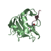
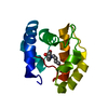
| 登録情報 | データベース: PDB / ID: 1ayb | ||||||
|---|---|---|---|---|---|---|---|
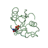
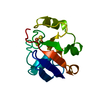
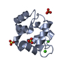
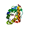
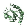
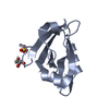
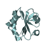
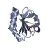
| タイトル | CRYSTAL STRUCTURES OF PEPTIDE COMPLEXES OF THE AMINO-TERMINAL SH2 DOMAIN OF THE SYP TYROSINE PHOSPHATASE | ||||||
 要素 要素 |
| ||||||
 キーワード キーワード | HYDROLASE(SH2 DOMAIN) | ||||||
| 機能・相同性 |  機能・相同性情報 機能・相同性情報IRS-mediated signalling / Signaling by ALK / IRS-related events triggered by IGF1R / PI-3K cascade:FGFR3 / PI-3K cascade:FGFR4 / Co-inhibition by BTLA / PI-3K cascade:FGFR1 / PI-3K cascade:FGFR2 / SOS-mediated signalling / FRS-mediated FGFR3 signaling ...IRS-mediated signalling / Signaling by ALK / IRS-related events triggered by IGF1R / PI-3K cascade:FGFR3 / PI-3K cascade:FGFR4 / Co-inhibition by BTLA / PI-3K cascade:FGFR1 / PI-3K cascade:FGFR2 / SOS-mediated signalling / FRS-mediated FGFR3 signaling / FRS-mediated FGFR4 signaling / MET activates PTPN11 / Regulation of IFNA/IFNB signaling / Signaling by LTK / FRS-mediated FGFR1 signaling / FRS-mediated FGFR2 signaling / Tie2 Signaling / PECAM1 interactions / PI3K/AKT activation / Interleukin-20 family signaling / Signaling by CSF3 (G-CSF) / GAB1 signalosome / PI3K Cascade / MAPK3 (ERK1) activation / MAPK1 (ERK2) activation / Co-inhibition by CTLA4 / Regulation of RUNX1 Expression and Activity / Interleukin-6 signaling / Negative regulation of FGFR3 signaling / Negative regulation of FGFR4 signaling / Downstream signal transduction / Negative regulation of FGFR1 signaling / Negative regulation of FGFR2 signaling / GPVI-mediated activation cascade / Interleukin-7 signaling / Spry regulation of FGF signaling / Co-inhibition by PD-1 / IRS activation / Signal attenuation / Activation of IRF3, IRF7 mediated by TBK1, IKKε (IKBKE) / Signaling by SCF-KIT / epithelial cell migration / positive regulation of signal transduction / negative regulation of hormone secretion / RAF/MAP kinase cascade / negative regulation of cortisol secretion / intestinal epithelial cell migration / microvillus organization / negative regulation of growth hormone secretion / PIP3 activates AKT signaling / genitalia development / Platelet sensitization by LDL / atrioventricular canal development / PI5P, PP2A and IER3 Regulate PI3K/AKT Signaling / negative regulation of cell adhesion mediated by integrin / Interleukin-3, Interleukin-5 and GM-CSF signaling / RET signaling / phosphatidylinositol 3-kinase activator activity / cerebellar cortex formation / negative regulation of neutrophil activation / positive regulation of hormone secretion / transmembrane receptor protein tyrosine kinase adaptor activity / regulation of protein export from nucleus / mammary gland development / positive regulation of lipopolysaccharide-mediated signaling pathway / positive regulation of ossification / cellular response to fatty acid / hormone metabolic process / negative regulation of chondrocyte differentiation / face morphogenesis / positive regulation of mesenchymal cell proliferation / platelet formation / triglyceride metabolic process / megakaryocyte development / negative regulation of type I interferon production / organ growth / peptide hormone receptor binding / neurotrophin TRK receptor signaling pathway / regulation of cell adhesion mediated by integrin / platelet-derived growth factor receptor signaling pathway / inner ear development / Bergmann glial cell differentiation / peptidyl-tyrosine dephosphorylation / non-membrane spanning protein tyrosine phosphatase activity / regulation of MAPK cascade / phosphoprotein phosphatase activity / positive regulation of epithelial cell migration / fibroblast growth factor receptor signaling pathway / protein localization to nucleus / positive regulation of insulin receptor signaling pathway / ephrin receptor signaling pathway / regulation of protein-containing complex assembly / phosphatidylinositol 3-kinase binding / lipid catabolic process / cell adhesion molecule binding / negative regulation of T cell proliferation / hormone-mediated signaling pathway / insulin-like growth factor receptor binding / phosphotyrosine residue binding / protein-tyrosine-phosphatase 類似検索 - 分子機能 | ||||||
| 生物種 |  | ||||||
| 手法 |  X線回折 / 解像度: 3 Å X線回折 / 解像度: 3 Å | ||||||
 データ登録者 データ登録者 | Lee, C.-H. / Kuriyan, J. | ||||||
 引用 引用 | ジャーナル: Structure / 年: 1994 タイトル: Crystal structures of peptide complexes of the amino-terminal SH2 domain of the Syp tyrosine phosphatase. 著者: Lee, C.H. / Kominos, D. / Jacques, S. / Margolis, B. / Schlessinger, J. / Shoelson, S.E. / Kuriyan, J. #1: ジャーナル: Cell(Cambridge,Mass.) / 年: 1993タイトル: Binding of a High Affinity Phosphotyrosyl Peptide to the Src Sh2 Domain: Crystal Structures of the Complexed and Peptide-Free Forms 著者: Waksman, G. / Shoelson, S.E. / Pant, N. / Cowburn, D. / Kuriyan, J. #2: ジャーナル: Curr.Opin.Struct.Biol. / 年: 1993タイトル: Structures of Sh2 and SH3 Domains 著者: Kuriyan, J. / Cowburn, D. #3: ジャーナル: Nature / 年: 1992タイトル: Crystal Structure of the Phosphotyrosine Recognition Domain Sh2 of V-Src Complexed with Tyrosine-Phosphorylated Peptides 著者: Waksman, G. / Kominos, D. / Robertson, S.R. / Pant, N. / Baltimore, D. / Birge, R.B. / Cowburn, D. / Hanafusa, H. / Mayer, B.J. / Overduin, M. / Resh, M.D. / Rios, C.B. / Silverman, L. / Kuriyan, J. | ||||||
| 履歴 |
|
- 構造の表示
構造の表示
| 構造ビューア | 分子: MolmilJmol/JSmol |
|---|
- ダウンロードとリンク
-ダウンロード
| PDBx/mmCIF形式 | 1ayb.cif.gz | 34.2 KB | 表示 | PDBx/mmCIF形式 |
|---|---|---|---|---|
| PDB形式 | pdb1ayb.ent.gz | 21.9 KB | 表示 | PDB形式 |
| PDBx/mmJSON形式 | 1ayb.json.gz | ツリー表示 | PDBx/mmJSON形式 | |
| その他 |  その他のダウンロード その他のダウンロード |
-検証レポート
| 文書・要旨 | 1ayb_validation.pdf.gz | 421.5 KB | 表示 | wwPDB検証レポート |
|---|---|---|---|---|
| 文書・詳細版 | 1ayb_full_validation.pdf.gz | 422.6 KB | 表示 | |
| XML形式データ | 1ayb_validation.xml.gz | 6.7 KB | 表示 | |
| CIF形式データ | 1ayb_validation.cif.gz | 7.9 KB | 表示 | |
| アーカイブディレクトリ | https://data.pdbj.org/pub/pdb/validation_reports/ay/1aybftp://data.pdbj.org/pub/pdb/validation_reports/ay/1ayb | HTTPS FTP |
-関連構造データ


-リンク
 PDBj
PDBj


- 集合体
集合体
| 登録構造単位 | 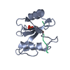
| ||||||||
|---|---|---|---|---|---|---|---|---|---|
| 1 |
| ||||||||
| 単位格子 |
|
-要素
| #1: タンパク質 | 分子量: 11528.979 Da / 分子数: 1 / 由来タイプ: 組換発現 / 由来: (組換発現) |
|---|---|
| #2: タンパク質・ペプチド | 分子量: 1378.335 Da / 分子数: 1 / 由来タイプ: 組換発現 / 由来: (組換発現) |
| Has protein modification | Y |
-実験情報
-実験
| 実験 | 手法: X線回折 |
|---|
- 試料調製
試料調製
| 結晶 | マシュー密度: 2.98 Å3/Da / 溶媒含有率: 58.72 % | ||||||||||||||||||||||||||||||||||||||||
|---|---|---|---|---|---|---|---|---|---|---|---|---|---|---|---|---|---|---|---|---|---|---|---|---|---|---|---|---|---|---|---|---|---|---|---|---|---|---|---|---|---|
| 結晶化 | *PLUS 温度: 4 ℃ / pH: 6 / 手法: 蒸気拡散法, ハンギングドロップ法 | ||||||||||||||||||||||||||||||||||||||||
| 溶液の組成 | *PLUS
|
-データ収集
| 放射 | 散乱光タイプ: x-ray |
|---|---|
| 放射波長 | 相対比: 1 |
| 反射 | *PLUS 最高解像度: 3 Å / 最低解像度: 30 Å / Num. all: 5965 / Num. obs: 2549 / % possible obs: 73 % / Rmerge(I) obs: 0.089 |
- 解析
解析
| ソフトウェア |
| ||||||||||||||||||||||||||||||||||||||||||||||||||||||||||||
|---|---|---|---|---|---|---|---|---|---|---|---|---|---|---|---|---|---|---|---|---|---|---|---|---|---|---|---|---|---|---|---|---|---|---|---|---|---|---|---|---|---|---|---|---|---|---|---|---|---|---|---|---|---|---|---|---|---|---|---|---|---|
| 精密化 | 解像度: 3→6 Å / σ(F): 2 /
| ||||||||||||||||||||||||||||||||||||||||||||||||||||||||||||
| 精密化ステップ | サイクル: LAST / 解像度: 3→6 Å
| ||||||||||||||||||||||||||||||||||||||||||||||||||||||||||||
| 拘束条件 |
| ||||||||||||||||||||||||||||||||||||||||||||||||||||||||||||
| ソフトウェア | *PLUS 名称: X-PLOR / 分類: refinement | ||||||||||||||||||||||||||||||||||||||||||||||||||||||||||||
| 精密化 | *PLUS Rfactor obs: 0.164 | ||||||||||||||||||||||||||||||||||||||||||||||||||||||||||||
| 溶媒の処理 | *PLUS | ||||||||||||||||||||||||||||||||||||||||||||||||||||||||||||
| 原子変位パラメータ | *PLUS | ||||||||||||||||||||||||||||||||||||||||||||||||||||||||||||
| 拘束条件 | *PLUS タイプ: x_angle_d / Dev ideal: 3 |
