 Movie
Movie Controller


+ Open data

- Basic information
| Entry | Database: PDB / ID: 1avz | ||||||
|---|---|---|---|---|---|---|---|
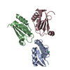
| Title | V-1 NEF PROTEIN IN COMPLEX WITH WILD TYPE FYN SH3 DOMAIN | ||||||
 Components Components |
| ||||||
 Keywords Keywords | COMPLEX (MYRISTYLATION/TRANSFERASE) / COMPLEX (MYRISTYLATION-TRANSFERASE) / GTP-BINDING / HIV-1 / PHOSPHORYLATION / NEF / SH3 DOMAIN / FYN / TYROSINE KINASE / COMPLEX (MYRISTYLATION-TRANSFERASE) complex | ||||||
| Function / homology |  Function and homology information Function and homology informationnegative regulation of hydrogen peroxide biosynthetic process / response to singlet oxygen / negative regulation of glycoprotein biosynthetic process / symbiont-mediated suppression of host antigen processing and presentation of peptide antigen via MHC class I / Reelin signalling pathway / symbiont-mediated suppression of host antigen processing and presentation of peptide antigen via MHC class II / perinuclear endoplasmic reticulum / NTRK2 activates RAC1 / symbiont-mediated suppression of host autophagy / regulation of glutamate receptor signaling pathway ...negative regulation of hydrogen peroxide biosynthetic process / response to singlet oxygen / negative regulation of glycoprotein biosynthetic process / symbiont-mediated suppression of host antigen processing and presentation of peptide antigen via MHC class I / Reelin signalling pathway / symbiont-mediated suppression of host antigen processing and presentation of peptide antigen via MHC class II / perinuclear endoplasmic reticulum / NTRK2 activates RAC1 / symbiont-mediated suppression of host autophagy / regulation of glutamate receptor signaling pathway / symbiont-mediated suppression of host apoptosis / Activated NTRK2 signals through FYN / reelin-mediated signaling pathway / SEMA3A-Plexin repulsion signaling by inhibiting Integrin adhesion / regulation of calcium ion import across plasma membrane / Platelet Adhesion to exposed collagen / cellular response to L-glutamate / CRMPs in Sema3A signaling / FLT3 signaling through SRC family kinases / regulation of T cell activation / G protein-coupled glutamate receptor signaling pathway / heart process / thioesterase binding / CD4 receptor binding / feeding behavior / positive regulation of protein localization to membrane / Nef and signal transduction / natural killer cell activation / Co-stimulation by CD28 / EPH-Ephrin signaling / DCC mediated attractive signaling / Nephrin family interactions / type 5 metabotropic glutamate receptor binding / CD28 dependent Vav1 pathway / Ephrin signaling / dendritic spine maintenance / negative regulation of dendritic spine maintenance / leukocyte migration / Regulation of KIT signaling / cellular response to peptide hormone stimulus / growth factor receptor binding / peptidase activator activity / phospholipase activator activity / tau-protein kinase activity / Co-inhibition by CTLA4 / EPHA-mediated growth cone collapse / negative regulation of T cell activation / Dectin-2 family / stimulatory C-type lectin receptor signaling pathway / peptide hormone receptor binding / Fc-gamma receptor signaling pathway involved in phagocytosis / PECAM1 interactions / CD8 receptor binding / response to amyloid-beta / FCGR activation / host cell Golgi membrane / Sema3A PAK dependent Axon repulsion / EPH-ephrin mediated repulsion of cells / Role of LAT2/NTAL/LAB on calcium mobilization / MHC class I protein binding / negative regulation of extrinsic apoptotic signaling pathway in absence of ligand / CD28 dependent PI3K/Akt signaling / regulation of calcium-mediated signaling / vascular endothelial growth factor receptor signaling pathway / cellular response to glycine / ephrin receptor signaling pathway / positive regulation of protein targeting to membrane / negative regulation of T cell receptor signaling pathway / glial cell projection / T cell costimulation / negative regulation of oxidative stress-induced intrinsic apoptotic signaling pathway / alpha-tubulin binding / ephrin receptor binding / postsynaptic density, intracellular component / phosphatidylinositol 3-kinase binding / GPVI-mediated activation cascade / T cell receptor binding / viral life cycle / phospholipase binding / Signaling by ERBB2 / negative regulation of angiogenesis / EPHB-mediated forward signaling / peptidyl-tyrosine phosphorylation / NCAM signaling for neurite out-growth / CD209 (DC-SIGN) signaling / FCGR3A-mediated IL10 synthesis / axon guidance / learning / cell surface receptor protein tyrosine kinase signaling pathway / Antigen activates B Cell Receptor (BCR) leading to generation of second messengers / Signaling by phosphorylated juxtamembrane, extracellular and kinase domain KIT mutants / Cell surface interactions at the vascular wall / non-specific protein-tyrosine kinase / FCGR3A-mediated phagocytosis / response to cocaine / actin filament / negative regulation of inflammatory response to antigenic stimulus / non-membrane spanning protein tyrosine kinase activity / Regulation of signaling by CBL / Signaling by SCF-KIT Similarity search - Function | ||||||
| Biological species |   Human immunodeficiency virus 1 Human immunodeficiency virus 1 Homo sapiens (human) Homo sapiens (human) | ||||||
| Method |  X-RAY DIFFRACTION / SYNCHROTRON / MOLECULAR REPLACEMENT / Resolution: 3 Å X-RAY DIFFRACTION / SYNCHROTRON / MOLECULAR REPLACEMENT / Resolution: 3 Å | ||||||
 Authors Authors | Arold, S. / Franken, P. / Dumas, C. | ||||||
 Citation Citation | Journal: Structure / Year: 1997 Title: The crystal structure of HIV-1 Nef protein bound to the Fyn kinase SH3 domain suggests a role for this complex in altered T cell receptor signaling. Authors: Arold, S. / Franken, P. / Strub, M.P. / Hoh, F. / Benichou, S. / Benarous, R. / Dumas, C. #1: Journal: Cell(Cambridge,Mass.) / Year: 1996Title: Crystal Structure of the Conserved Core of HIV-1 Nef Complexed with a Src Family SH3 Domain Authors: Lee, C.H. / Saksela, K. / Mirza, U.A. / Chait, B.T. / Kuriyan, J. #2: Journal: Nat.Struct.Biol. / Year: 1996Title: The Solution Structure of HIV-1 Nef Reveals an Unexpected Fold and Permits Delineation of the Binding Surface for the SH3 Domain of HCK Tyrosine Protein Kinase Authors: Grzesiek, S. / Bax, A. / Clore, G.M. / Gronenborn, A.M. / Hu, J.S. / Kaufman, J. / Palmer, I. / Stahl, S.J. / Wingfield, P.T. | ||||||
| History |
|
- Structure visualization
| Structure viewer | Molecule: MolmilJmol/JSmol |
|---|
- Downloads & links
-Download
| PDBx/mmCIF format | 1avz.cif.gz | 62 KB | Display | PDBx/mmCIF format |
|---|---|---|---|---|
| PDB format | pdb1avz.ent.gz | 46.1 KB | Display | PDB format |
| PDBx/mmJSON format | 1avz.json.gz | Tree view | PDBx/mmJSON format | |
| Others |  Other downloads Other downloads |
-Validation report
| Arichive directory | https://data.pdbj.org/pub/pdb/validation_reports/av/1avzftp://data.pdbj.org/pub/pdb/validation_reports/av/1avz | HTTPS FTP |
|---|
-Related structure data
| Related structure data |  1avvC  1efnS  1shfS S: Starting model for refinement C: citing same article ( |
|---|---|
| Similar structure data |



-Links
 PDBj
PDBj















- Assembly
| Deposited unit | 
| ||||||||
|---|---|---|---|---|---|---|---|---|---|
| 1 |
| ||||||||
| Unit cell |
|
-Components
| #1: Protein | Mass: 17568.723 Da / Num. of mol.: 2 / Fragment: CONSERVED CORE DOMAIN Mutation: N-TERMINAL RESIDUES GS (PART OF A THROMBIN CLEAVAGE SITE) Source method: isolated from a genetically manipulated source Source: (gene. exp.) Human immunodeficiency virus 1 / Genus: Lentivirus / Cell line: BL21 / Gene: HIV-1 NEF / Variant: LAI ISOLATE / Plasmid: PGEX-2T / Species (production host): Escherichia coli / Production host:   Escherichia coli BL21(DE3) (bacteria) / Strain (production host): BL21 (DE3) / References: UniProt: P03406 Escherichia coli BL21(DE3) (bacteria) / Strain (production host): BL21 (DE3) / References: UniProt: P03406#2: Protein | | Mass: 6524.066 Da / Num. of mol.: 1 / Fragment: SRC-HOMOLOGY 3 DOMAIN Source method: isolated from a genetically manipulated source Source: (gene. exp.) Homo sapiens (human) / Cell line: BL21 / Gene: FYN TYROSINE KINASE / Plasmid: PGEX-2T / Species (production host): Escherichia coli / Production host: Escherichia coli BL21(DE3) (bacteria) / Strain (production host): BL21 (DE3) / References: UniProt: P06241, EC: 2.7.1.112 |
|---|
-Experimental details
-Experiment
| Experiment | Method: X-RAY DIFFRACTION / Number of used crystals: 1 |
|---|
- Sample preparation
| Crystal | Density Matthews: 4.2 Å3/Da / Density % sol: 70 % |
|---|---|
| Crystal grow | pH: 8.9 / Details: pH 8.9 |
| Crystal | *PLUS |
| Crystal grow | *PLUS Method: unknown / Details: unpublished data |
-Data collection
| Diffraction | Mean temperature: 100 K |
|---|---|
| Diffraction source | Source: SYNCHROTRON / Site: ESRF  / Beamline: BM02 / Wavelength: 1.0373 / Beamline: BM02 / Wavelength: 1.0373 |
| Detector | Type: PRINCETON 2K / Detector: CCD / Date: Oct 4, 1996 / Details: TWO BENT MIRRORS |
| Radiation | Monochromator: SI(111) / Monochromatic (M) / Laue (L): M / Scattering type: x-ray |
| Radiation wavelength | Wavelength: 1.0373 Å / Relative weight: 1 |
| Reflection | Resolution: 3→25 Å / Num. obs: 14778 / % possible obs: 91 % / Observed criterion σ(I): 2 / Redundancy: 4.1 % / Rmerge(I) obs: 0.067 |
| Reflection | *PLUS Num. measured all: 57175 |
- Processing
| Software |
| ||||||||||||||||||||||||||||||||||||||||||||||||||||||||||||
|---|---|---|---|---|---|---|---|---|---|---|---|---|---|---|---|---|---|---|---|---|---|---|---|---|---|---|---|---|---|---|---|---|---|---|---|---|---|---|---|---|---|---|---|---|---|---|---|---|---|---|---|---|---|---|---|---|---|---|---|---|---|
| Refinement | Method to determine structure: MOLECULAR REPLACEMENT Starting model: PDB ENTRIES 1EFN AND 1SHF Resolution: 3→30 Å / Data cutoff high absF: 10000000 / Data cutoff low absF: 0.001 / σ(F): 2
| ||||||||||||||||||||||||||||||||||||||||||||||||||||||||||||
| Displacement parameters | Biso mean: 33 Å2 | ||||||||||||||||||||||||||||||||||||||||||||||||||||||||||||
| Refinement step | Cycle: LAST / Resolution: 3→30 Å
| ||||||||||||||||||||||||||||||||||||||||||||||||||||||||||||
| Refine LS restraints |
| ||||||||||||||||||||||||||||||||||||||||||||||||||||||||||||
| Software | *PLUS Name: X-PLOR / Version: 3.8 / Classification: refinement | ||||||||||||||||||||||||||||||||||||||||||||||||||||||||||||
| Refine LS restraints | *PLUS
|
