 Movie
Movie Controller

[English] 日本語
 Yorodumi
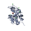
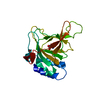
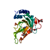
- PDB-1avp: STRUCTURE OF HUMAN ADENOVIRUS 2 PROTEINASE WITH ITS 11 AMINO ACID... -
+ Open data

- Basic information
| Entry | Database: PDB / ID: 1avp | ||||||
|---|---|---|---|---|---|---|---|
| Title | STRUCTURE OF HUMAN ADENOVIRUS 2 PROTEINASE WITH ITS 11 AMINO ACID COFACTOR | ||||||
 Components Components | (ADENOVIRAL PROTEINASE) x 2 | ||||||
 Keywords Keywords | HYDROLASE / THIOL HYDROLASE / VIRAL PROTEINASE / PEPTIDE COFACTOR | ||||||
| Function / homology |  Function and homology information Function and homology informationadenain / nuclear capsid assembly / lysis of host organelle involved in viral entry into host cell / viral procapsid / microtubule-dependent intracellular transport of viral material towards nucleus / viral release from host cell / cysteine-type peptidase activity / virion component / host cell / viral capsid ...adenain / nuclear capsid assembly / lysis of host organelle involved in viral entry into host cell / viral procapsid / microtubule-dependent intracellular transport of viral material towards nucleus / viral release from host cell / cysteine-type peptidase activity / virion component / host cell / viral capsid / host cell cytoplasm / cysteine-type endopeptidase activity / host cell nucleus / proteolysis / DNA binding Similarity search - Function | ||||||
| Biological species |   Human adenovirus 2Human adenovirus C Human adenovirus 2Human adenovirus C | ||||||
| Method |  X-RAY DIFFRACTION / SYNCHROTRON / SIRAS SOFTWARE USED : NULL STARTING MODEL FOR MOLECULAR REPLACEMENT: NULL / Resolution: 2.6 Å X-RAY DIFFRACTION / SYNCHROTRON / SIRAS SOFTWARE USED : NULL STARTING MODEL FOR MOLECULAR REPLACEMENT: NULL / Resolution: 2.6 Å | ||||||
 Authors Authors | Ding, J. / Mcgrath, W.J. / Sweet, R.M. / Mangel, W.F. | ||||||
 Citation Citation | Journal: EMBO J. / Year: 1996 Title: Crystal structure of the human adenovirus proteinase with its 11 amino acid cofactor. Authors: Ding, J. / McGrath, W.J. / Sweet, R.M. / Mangel, W.F. #1: Journal: To be PublishedTitle: Characterization of the Human Adenovirus Proteinase Activity in Disrupted Virus Particles Authors: Mcgrath, W.J. / Abola, A.P. / Toledo, D.L. / Brown, M.T. / Mangel, W.F. #2: Journal: J.Biol.Chem. / Year: 1996Title: Characterization of Three Components of Human Adenovirus Activity in Vitro Authors: Mangel, W.F. / Toledo, D.L. / Brown, M.T. / Martin, J.H. / Mcgrath, W.J. #3: Journal: Nature / Year: 1993Title: Viral DNA and a Viral Peptide Can Act as Cofactors of Adenovirus Virion Proteinase Activity Authors: Mangel, W.F. / Mcgrath, W.J. / Toledo, D.L. / Anderson, C.W. | ||||||
| History |
| ||||||
| Remark 700 | SHEET SHEET SHEET_ID: (), DETERMINATION METHOD: PROCHECK. |
- Structure visualization
| Structure viewer | Molecule: MolmilJmol/JSmol |
|---|
- Downloads & links
-Download
| PDBx/mmCIF format | 1avp.cif.gz | 62.8 KB | Display | PDBx/mmCIF format |
|---|---|---|---|---|
| PDB format | pdb1avp.ent.gz | 50.5 KB | Display | PDB format |
| PDBx/mmJSON format | 1avp.json.gz | Tree view | PDBx/mmJSON format | |
| Others |  Other downloads Other downloads |
-Validation report
| Summary document | 1avp_validation.pdf.gz | 431.3 KB | Display | wwPDB validaton report |
|---|---|---|---|---|
| Full document | 1avp_full_validation.pdf.gz | 439.8 KB | Display | |
| Data in XML | 1avp_validation.xml.gz | 11.9 KB | Display | |
| Data in CIF | 1avp_validation.cif.gz | 15.2 KB | Display | |
| Arichive directory | https://data.pdbj.org/pub/pdb/validation_reports/av/1avpftp://data.pdbj.org/pub/pdb/validation_reports/av/1avp | HTTPS FTP |
-Related structure data
| Similar structure data |
|---|

-Links
 PDBj
PDBj
- Assembly
| Deposited unit | 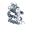
| ||||||||
|---|---|---|---|---|---|---|---|---|---|
| 1 |
| ||||||||
| Unit cell |
|
-Components
| #1: Protein | Mass: 23114.350 Da / Num. of mol.: 1 / Fragment: MAIN Source method: isolated from a genetically manipulated source Details: PVIC PEPTIDE COFACTOR DISULFIDE BONDS TO CYS104 OF AVP Source: (gene. exp.) Human adenovirus 2 / Genus: Mastadenovirus / Species: Human adenovirus C / Fragment: MAIN / Plasmid: PET 13 / Species (production host): Escherichia coli / Production host:  |
|---|---|
| #2: Protein/peptide | Mass: 1353.640 Da / Num. of mol.: 1 / Fragment: PEPT / Source method: obtained synthetically Details: PVIC PEPTIDE IS CHEMICALLY SYNTHESIZED PVIC PEPTIDE COFACTOR DISULFIDE BONDS TO CYS104 OF AVP Source: (synth.) Human adenovirus C / References: UniProt: P03274 |
| #3: Water | ChemComp-HOH /  Mass: 18.015 Da / Num. of mol.: 45 / Source method: isolated from a natural source / Formula: H2O Mass: 18.015 Da / Num. of mol.: 45 / Source method: isolated from a natural source / Formula: H2O |
-Experimental details
-Experiment
| Experiment | Method: X-RAY DIFFRACTION / Number of used crystals: 4 |
|---|
- Sample preparation
| Crystal | Density Matthews: 3.85 Å3/Da / Density % sol: 68.06 % | ||||||||||||||||||
|---|---|---|---|---|---|---|---|---|---|---|---|---|---|---|---|---|---|---|---|
| Crystal grow | pH: 6.5 / Details: pH 6.5 | ||||||||||||||||||
| Crystal | *PLUS Density % sol: 68 % | ||||||||||||||||||
| Crystal grow | *PLUS Method: vapor diffusion, hanging drop / Details: McGrath, W.J., (1996) J. Struct. Biol., 117, 77. / pH: 7.5 | ||||||||||||||||||
| Components of the solutions | *PLUS
|
-Data collection
| Diffraction | Mean temperature: 300 K |
|---|---|
| Diffraction source | Source: SYNCHROTRON / Site: NSLS  / Beamline: X12C / Wavelength: 1.15 / Beamline: X12C / Wavelength: 1.15 |
| Detector | Type: ENRAF-NONIUS FAST / Detector: DIFFRACTOMETER / Date: May 10, 1995 / Details: COLLIMATOR |
| Radiation | Monochromator: SI(111) / Monochromatic (M) / Laue (L): M / Scattering type: x-ray |
| Radiation wavelength | Wavelength: 1.15 Å / Relative weight: 1 |
| Reflection | Resolution: 2.6→50 Å / Num. obs: 11559 / % possible obs: 98.8 % / Observed criterion σ(I): 3 / Redundancy: 5 % / Rmerge(I) obs: 0.077 / Net I/σ(I): 20 |
| Reflection shell | Resolution: 2.6→2.69 Å / Redundancy: 6 % / Rmerge(I) obs: 0.145 / Mean I/σ(I) obs: 16 / % possible all: 97.6 |
- Processing
| Software |
| ||||||||||||||||||||||||||||||||||||||||||||||||||||||||||||
|---|---|---|---|---|---|---|---|---|---|---|---|---|---|---|---|---|---|---|---|---|---|---|---|---|---|---|---|---|---|---|---|---|---|---|---|---|---|---|---|---|---|---|---|---|---|---|---|---|---|---|---|---|---|---|---|---|---|---|---|---|---|
| Refinement | Method to determine structure: SIRAS SOFTWARE USED : NULL STARTING MODEL FOR MOLECULAR REPLACEMENT: NULL Resolution: 2.6→6 Å / Rfactor Rfree error: 0.007 / Cross valid method: THROUGHOUT
| ||||||||||||||||||||||||||||||||||||||||||||||||||||||||||||
| Refine analyze | Luzzati coordinate error obs: 0.1 Å / Luzzati d res low obs: 6 Å | ||||||||||||||||||||||||||||||||||||||||||||||||||||||||||||
| Refinement step | Cycle: LAST / Resolution: 2.6→6 Å
| ||||||||||||||||||||||||||||||||||||||||||||||||||||||||||||
| Refine LS restraints |
| ||||||||||||||||||||||||||||||||||||||||||||||||||||||||||||
| LS refinement shell | Resolution: 2.6→2.64 Å / Rfactor Rfree error: 0.053 /
| ||||||||||||||||||||||||||||||||||||||||||||||||||||||||||||
| Xplor file |
| ||||||||||||||||||||||||||||||||||||||||||||||||||||||||||||
| Software | *PLUS Name: X-PLOR / Classification: refinement | ||||||||||||||||||||||||||||||||||||||||||||||||||||||||||||
| Refine LS restraints | *PLUS
|
