 Movie
Movie Controller


+ Open data

- Basic information
| Entry | Database: PDB / ID: 1al3 | ||||||
|---|---|---|---|---|---|---|---|
| Title | COFACTOR BINDING FRAGMENT OF CYSB FROM KLEBSIELLA AEROGENES | ||||||
 Components Components | CYS REGULON TRANSCRIPTIONAL ACTIVATOR CYSB | ||||||
 Keywords Keywords | TRANSCRIPTION REGULATION / LYSR FAMILY / CYSTEINE BIOSYNTHESIS | ||||||
| Function / homology |  Function and homology information Function and homology informationL-cysteine biosynthetic process / transcription cis-regulatory region binding / DNA-binding transcription factor activity / cytoplasm Similarity search - Function | ||||||
| Biological species |  Klebsiella aerogenes (bacteria) Klebsiella aerogenes (bacteria) | ||||||
| Method |  X-RAY DIFFRACTION / SYNCHROTRON / MIR / Resolution: 1.8 Å X-RAY DIFFRACTION / SYNCHROTRON / MIR / Resolution: 1.8 Å | ||||||
 Authors Authors | Verschueren, K.H.G. / Tyrrell, R. / Wilkinson, A.J. | ||||||
 Citation Citation | Journal: Structure / Year: 1997 Title: The structure of the cofactor-binding fragment of the LysR family member, CysB: a familiar fold with a surprising subunit arrangement. Authors: Tyrrell, R. / Verschueren, K.H. / Dodson, E.J. / Murshudov, G.N. / Addy, C. / Wilkinson, A.J. | ||||||
| History |
|
- Structure visualization
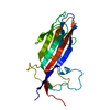
| Structure viewer | Molecule: MolmilJmol/JSmol |
|---|
- Downloads & links
-Download
| PDBx/mmCIF format | 1al3.cif.gz | 70.8 KB | Display | PDBx/mmCIF format |
|---|---|---|---|---|
| PDB format | pdb1al3.ent.gz | 52.5 KB | Display | PDB format |
| PDBx/mmJSON format | 1al3.json.gz | Tree view | PDBx/mmJSON format | |
| Others |  Other downloads Other downloads |
-Validation report
| Arichive directory | https://data.pdbj.org/pub/pdb/validation_reports/al/1al3ftp://data.pdbj.org/pub/pdb/validation_reports/al/1al3 | HTTPS FTP |
|---|
-Related structure data
| Similar structure data |
|---|


-Links
 PDBj
PDBj- Assembly
| Deposited unit | 
| ||||||||
|---|---|---|---|---|---|---|---|---|---|
| 1 |
| ||||||||
| Unit cell |
| ||||||||
| Components on special symmetry positions |
|
-Components
| #1: Protein | Mass: 36008.109 Da / Num. of mol.: 1 / Fragment: COFACTOR BINDING FRAGMENT, RESIDUES 88 - 324 Source method: isolated from a genetically manipulated source Source: (gene. exp.) Klebsiella aerogenes (bacteria)Description: KLEBSIELLA AEROGENES NCTC 418. CHYMOTRYPTIC C-TERMINAL COFACTOR BINDING FRAGMENT OF CYSB Gene: CYSB / Plasmid: PKK223-3 / Production host: |
|---|---|
| #2: Chemical | ChemComp-SO4 /   Mass: 96.063 Da / Num. of mol.: 1 / Source method: obtained synthetically / Formula: SO4 Mass: 96.063 Da / Num. of mol.: 1 / Source method: obtained synthetically / Formula: SO4 |
| #3: Water | ChemComp-HOH /  Mass: 18.015 Da / Num. of mol.: 253 / Source method: isolated from a natural source / Formula: H2O Mass: 18.015 Da / Num. of mol.: 253 / Source method: isolated from a natural source / Formula: H2O |
-Experimental details
-Experiment
| Experiment | Method: X-RAY DIFFRACTION / Number of used crystals: 1 |
|---|
- Sample preparation
| Crystal | Density Matthews: 2.22 Å3/Da / Density % sol: 44 % | |||||||||||||||
|---|---|---|---|---|---|---|---|---|---|---|---|---|---|---|---|---|
| Crystal grow | Temperature: 291 K / Method: vapor diffusion, hanging drop / pH: 6.5 Details: COFACTOR BINDING DOMAIN WAS CRYSTALLISED IN HANGING DROPS AT 18 DEGREES CELSIUS FROM 14% MONO-METHYLETHER PEG750, 100MM MES PH 6.5, vapor diffusion - hanging drop, temperature 291K | |||||||||||||||
| Crystal grow | *PLUS Method: vapor diffusion, hanging drop | |||||||||||||||
| Components of the solutions | *PLUS
|
-Data collection
| Diffraction | Mean temperature: 293 K |
|---|---|
| Diffraction source | Source: SYNCHROTRON / Site: EMBL/DESY, HAMBURG  / Beamline: X11 / Wavelength: 0.91 / Beamline: X11 / Wavelength: 0.91 |
| Detector | Type: MAR scanner 300 mm plate / Detector: IMAGE PLATE / Date: Dec 1, 1994 |
| Radiation | Monochromatic (M) / Laue (L): M / Scattering type: x-ray |
| Radiation wavelength | Wavelength: 0.91 Å / Relative weight: 1 |
| Reflection | Resolution: 1.8→20 Å / Num. obs: 22479 / % possible obs: 94.1 % / Observed criterion σ(I): 0 / Redundancy: 2.5 % / Rmerge(I) obs: 0.044 / Net I/σ(I): 7.7 |
| Reflection shell | Resolution: 1.8→1.9 Å / Redundancy: 2.1 % / Rmerge(I) obs: 0.319 / Mean I/σ(I) obs: 1.6 / % possible all: 84.9 |
| Reflection | *PLUS Num. obs: 20911 / % possible obs: 93 % |
| Reflection shell | *PLUS % possible obs: 84.9 % |
- Processing
| Software |
| ||||||||||||||||||||||||||||||||||||||||||||||||||||||||||||||||||||||||||||||||||||
|---|---|---|---|---|---|---|---|---|---|---|---|---|---|---|---|---|---|---|---|---|---|---|---|---|---|---|---|---|---|---|---|---|---|---|---|---|---|---|---|---|---|---|---|---|---|---|---|---|---|---|---|---|---|---|---|---|---|---|---|---|---|---|---|---|---|---|---|---|---|---|---|---|---|---|---|---|---|---|---|---|---|---|---|---|---|
| Refinement | Method to determine structure: MIR / Resolution: 1.8→20 Å / Cross valid method: THROUGHOUT
| ||||||||||||||||||||||||||||||||||||||||||||||||||||||||||||||||||||||||||||||||||||
| Displacement parameters | Biso mean: 29.8 Å2 | ||||||||||||||||||||||||||||||||||||||||||||||||||||||||||||||||||||||||||||||||||||
| Refine analyze | Luzzati sigma a obs: 0.15 Å | ||||||||||||||||||||||||||||||||||||||||||||||||||||||||||||||||||||||||||||||||||||
| Refinement step | Cycle: LAST / Resolution: 1.8→20 Å
| ||||||||||||||||||||||||||||||||||||||||||||||||||||||||||||||||||||||||||||||||||||
| Refine LS restraints |
| ||||||||||||||||||||||||||||||||||||||||||||||||||||||||||||||||||||||||||||||||||||
| Software | *PLUS Name: REFMAC / Classification: refinement | ||||||||||||||||||||||||||||||||||||||||||||||||||||||||||||||||||||||||||||||||||||
| Refinement | *PLUS Num. reflection obs: 20911 / Rfactor obs: 0.179 | ||||||||||||||||||||||||||||||||||||||||||||||||||||||||||||||||||||||||||||||||||||
| Solvent computation | *PLUS | ||||||||||||||||||||||||||||||||||||||||||||||||||||||||||||||||||||||||||||||||||||
| Displacement parameters | *PLUS | ||||||||||||||||||||||||||||||||||||||||||||||||||||||||||||||||||||||||||||||||||||
| Refine LS restraints | *PLUS Type: p_bond_d / Dev ideal: 0.014 |
