 Movie
Movie Controller

[English] 日本語
 Yorodumi
- PDB-4lon: Crystal structure of monomeric sulphate free form of ligand bindi... -
+ Open data

- Basic information
| Entry | Database: PDB / ID: 4lon | ||||||
|---|---|---|---|---|---|---|---|
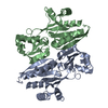
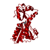
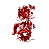
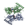
| Title | Crystal structure of monomeric sulphate free form of ligand binding domain of CysB,an LTTR from Salmonella typhimurium LT2 | ||||||
 Components Components | HTH-type transcriptional regulator CysB | ||||||
 Keywords Keywords | GENE REGULATION / LTTR / CysB / wHTH motif / alpha/beta fold / Rossmann fold / Transcription regulation / O-acetyl serine / N-acetyl serine binding / DNA binding | ||||||
| Function / homology |  Function and homology information Function and homology informationL-cysteine biosynthetic process / transcription cis-regulatory region binding / DNA-binding transcription factor activity / regulation of DNA-templated transcription / cytoplasm Similarity search - Function | ||||||
| Biological species |  Salmonella enterica subsp. enterica serovar Typhimurium (bacteria) Salmonella enterica subsp. enterica serovar Typhimurium (bacteria) | ||||||
| Method |  X-RAY DIFFRACTION / SYNCHROTRON / MOLECULAR REPLACEMENT / Resolution: 2.2 Å X-RAY DIFFRACTION / SYNCHROTRON / MOLECULAR REPLACEMENT / Resolution: 2.2 Å | ||||||
 Authors Authors | Mittal, M. / Singh, A.K. / Kumaran, S. | ||||||
 Citation Citation | Journal: To be Published Title: Crystal structure of monomeric sulphate free form of ligand binding domain of CysB,an LTTR from Salmonella typhimurium LT2 Authors: Mittal, M. / Singh, A.K. / Kumaran, S. | ||||||
| History |
|
- Structure visualization
| Structure viewer | Molecule: MolmilJmol/JSmol |
|---|
- Downloads & links
-Download
| PDBx/mmCIF format | 4lon.cif.gz | 59.8 KB | Display | PDBx/mmCIF format |
|---|---|---|---|---|
| PDB format | pdb4lon.ent.gz | 42.7 KB | Display | PDB format |
| PDBx/mmJSON format | 4lon.json.gz | Tree view | PDBx/mmJSON format | |
| Others |  Other downloads Other downloads |
-Validation report
| Arichive directory | https://data.pdbj.org/pub/pdb/validation_reports/lo/4lonftp://data.pdbj.org/pub/pdb/validation_reports/lo/4lon | HTTPS FTP |
|---|
-Related structure data
| Related structure data | 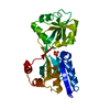 1al3S S: Starting model for refinement |
|---|---|
| Similar structure data |


-Links
 PDBj
PDBj- Assembly
| Deposited unit | 
| ||||||||
|---|---|---|---|---|---|---|---|---|---|
| 1 | 
| ||||||||
| Unit cell |
|
-Components
| #1: Protein | Mass: 26689.342 Da / Num. of mol.: 1 / Fragment: UNP residues 86-324 Source method: isolated from a genetically manipulated source Details: IPTG Inducible Source: (gene. exp.) Salmonella enterica subsp. enterica serovar Typhimurium (bacteria)Strain: LT2 / SGSC1412 / ATCC 700720 / Gene: cysB, STM1713 / Plasmid: Pet28a / Production host: |
|---|---|
| #2: Water | ChemComp-HOH /  Mass: 18.015 Da / Num. of mol.: 44 / Source method: isolated from a natural source / Formula: H2O Mass: 18.015 Da / Num. of mol.: 44 / Source method: isolated from a natural source / Formula: H2O |
-Experimental details
-Experiment
| Experiment | Method: X-RAY DIFFRACTION / Number of used crystals: 1 |
|---|
- Sample preparation
| Crystal | Density Matthews: 1.98 Å3/Da / Density % sol: 37.8 % |
|---|---|
| Crystal grow | Temperature: 291 K / Method: vapor diffusion, sitting drop / pH: 6.5 Details: 20%(w/v) PEG8000, 0.1M Sodium cacodylate pH 6.5, 0.2M magnesium acetate, VAPOR DIFFUSION, SITTING DROP, temperature 291K |
-Data collection
| Diffraction | Mean temperature: 100 K |
|---|---|
| Diffraction source | Source: SYNCHROTRON / Site: ESRF  / Beamline: BM14 / Wavelength: 0.97755 Å / Beamline: BM14 / Wavelength: 0.97755 Å |
| Detector | Type: MARRESEARCH / Detector: CCD / Date: May 24, 2012 / Details: mirrors |
| Radiation | Monochromator: Chanel cut Si111 / Protocol: SINGLE WAVELENGTH / Monochromatic (M) / Laue (L): M / Scattering type: x-ray |
| Radiation wavelength | Wavelength: 0.97755 Å / Relative weight: 1 |
| Reflection | Resolution: 2.2→50 Å / Num. all: 70402 / Num. obs: 70402 / % possible obs: 99.7 % / Observed criterion σ(F): 2 / Observed criterion σ(I): 2 / Redundancy: 5.8 % / Rmerge(I) obs: 0.072 / Net I/σ(I): 24.542 |
- Processing
| Software |
| ||||||||||||||||||||||||||||||
|---|---|---|---|---|---|---|---|---|---|---|---|---|---|---|---|---|---|---|---|---|---|---|---|---|---|---|---|---|---|---|---|
| Refinement | Method to determine structure: MOLECULAR REPLACEMENT Starting model: 1AL3 Resolution: 2.2→44.235 Å / SU ML: 0.17 / Isotropic thermal model: Isotropic / σ(F): 1.36 / Phase error: 29.86 / Stereochemistry target values: ML
| ||||||||||||||||||||||||||||||
| Solvent computation | Shrinkage radii: 0.86 Å / VDW probe radii: 1.1 Å / Solvent model: FLAT BULK SOLVENT MODEL / Bsol: 35.098 Å2 / ksol: 0.317 e/Å3 | ||||||||||||||||||||||||||||||
| Displacement parameters |
| ||||||||||||||||||||||||||||||
| Refinement step | Cycle: LAST / Resolution: 2.2→44.235 Å
| ||||||||||||||||||||||||||||||
| Refine LS restraints |
| ||||||||||||||||||||||||||||||
| LS refinement shell | Refine-ID: X-RAY DIFFRACTION / Total num. of bins used: 4
|
