 Movie
Movie Controller


+ Open data

- Basic information
| Entry | Database: PDB / ID: 1akh | ||||||
|---|---|---|---|---|---|---|---|
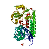
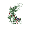
| Title | MAT A1/ALPHA2/DNA TERNARY COMPLEX | ||||||
 Components Components |
| ||||||
 Keywords Keywords | DNA BINDING PROTEIN/DNA / COMPLEX (TWO DNA-BINDING PROTEINS-DNA) / COMPLEX / DNA-BINDING PROTEIN / DNA / TRANSCRIPTION REGULATION / DNA BINDING PROTEIN-DNA COMPLEX | ||||||
| Function / homology |  Function and homology information Function and homology information: / regulation of mating-type specific transcription, DNA-templated / RNA polymerase II transcription repressor complex / DNA binding, bending / DNA-binding transcription repressor activity, RNA polymerase II-specific / sequence-specific DNA binding / DNA-binding transcription factor activity, RNA polymerase II-specific / RNA polymerase II cis-regulatory region sequence-specific DNA binding / regulation of transcription by RNA polymerase II / negative regulation of transcription by RNA polymerase II ...: / regulation of mating-type specific transcription, DNA-templated / RNA polymerase II transcription repressor complex / DNA binding, bending / DNA-binding transcription repressor activity, RNA polymerase II-specific / sequence-specific DNA binding / DNA-binding transcription factor activity, RNA polymerase II-specific / RNA polymerase II cis-regulatory region sequence-specific DNA binding / regulation of transcription by RNA polymerase II / negative regulation of transcription by RNA polymerase II / DNA binding / nucleus Similarity search - Function | ||||||
| Biological species |  | ||||||
| Method |  X-RAY DIFFRACTION / MOLECULAR REPLACEMENT / Resolution: 2.5 Å X-RAY DIFFRACTION / MOLECULAR REPLACEMENT / Resolution: 2.5 Å | ||||||
 Authors Authors | Li, T. / Jin, Y. / Vershon, A.K. / Wolberger, C. | ||||||
 Citation Citation | Journal: Nucleic Acids Res. / Year: 1998 Title: Crystal structure of the MATa1/MATalpha2 homeodomain heterodimer in complex with DNA containing an A-tract. Authors: Li, T. / Jin, Y. / Vershon, A.K. / Wolberger, C. #1: Journal: Proteins / Year: 1995Title: Crystallization and Preliminary X-Ray Diffraction Studies of an A1/Alpha 2/DNA Ternary Complex Authors: Li, T. / Stark, M. / Johnson, A.D. / Wolberger, C. #2: Journal: Science / Year: 1995Title: Crystal Structure of the MATa1/MAT Alpha 2 Homeodomain Heterodimer Bound to DNA Authors: Li, T. / Stark, M.R. / Johnson, A.D. / Wolberger, C. #3: Journal: Science / Year: 1995Title: Erratum. Crystal Structure of the MATa1/MAT Alpha 2 Homeodomain Heterodimer Bound to DNA Authors: Li, T. / Stark, M.R. / Johnson, A.D. / Wolberger, C. | ||||||
| History |
|
- Structure visualization
| Structure viewer | Molecule: MolmilJmol/JSmol |
|---|
- Downloads & links
-Download
| PDBx/mmCIF format | 1akh.cif.gz | 58.2 KB | Display | PDBx/mmCIF format |
|---|---|---|---|---|
| PDB format | pdb1akh.ent.gz | 43.3 KB | Display | PDB format |
| PDBx/mmJSON format | 1akh.json.gz | Tree view | PDBx/mmJSON format | |
| Others |  Other downloads Other downloads |
-Validation report
| Arichive directory | https://data.pdbj.org/pub/pdb/validation_reports/ak/1akhftp://data.pdbj.org/pub/pdb/validation_reports/ak/1akh | HTTPS FTP |
|---|
-Related structure data
| Related structure data |  1yrnS S: Starting model for refinement |
|---|---|
| Similar structure data |


-Links
 PDBj
PDBj


- Assembly
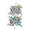
| Deposited unit | 
| ||||||||
|---|---|---|---|---|---|---|---|---|---|
| 1 |
| ||||||||
| Unit cell |
|
-Components
| #1: DNA chain | Mass: 6413.210 Da / Num. of mol.: 1 / Source method: obtained synthetically |
|---|---|
| #2: DNA chain | Mass: 6466.217 Da / Num. of mol.: 1 / Source method: obtained synthetically |
| #3: Protein | Mass: 7165.479 Da / Num. of mol.: 1 Source method: isolated from a genetically manipulated source Source: (gene. exp.) Gene: MAT A1 RESIDUES 66 - 126 / Plasmid: PMS-K66 / Gene (production host): MAT A1 RESIDUES 66 - 126 / Production host:  |
| #4: Protein | Mass: 9773.306 Da / Num. of mol.: 1 Source method: isolated from a genetically manipulated source Source: (gene. exp.) Gene: MAT ALPHA2 RESIDUES 128 - 210 / Plasmid: PAV105 / Gene (production host): MAT ALPHA2 RESIDUES 128 - 210 / Production host: |
| #5: Water | ChemComp-HOH /  Mass: 18.015 Da / Num. of mol.: 51 / Source method: isolated from a natural source / Formula: H2O Mass: 18.015 Da / Num. of mol.: 51 / Source method: isolated from a natural source / Formula: H2O |
-Experimental details
-Experiment
| Experiment | Method: X-RAY DIFFRACTION / Number of used crystals: 3 |
|---|
- Sample preparation
| Crystal | Density Matthews: 3.78 Å3/Da / Density % sol: 67.6 % | ||||||||||||||||||||||||||||||||||||||||||||||||
|---|---|---|---|---|---|---|---|---|---|---|---|---|---|---|---|---|---|---|---|---|---|---|---|---|---|---|---|---|---|---|---|---|---|---|---|---|---|---|---|---|---|---|---|---|---|---|---|---|---|
| Crystal grow | Temperature: 295 K / Method: vapor diffusion, hanging drop / pH: 7 Details: 100MM HEPES PH 7.0, 20MM CACL2, 5MM [CO(NH3)6]CL3., VAPOR DIFFUSION, HANGING DROP, temperature 295.00K | ||||||||||||||||||||||||||||||||||||||||||||||||
| Components of the solutions |
| ||||||||||||||||||||||||||||||||||||||||||||||||
| Crystal | *PLUS Density % sol: 67.6 % | ||||||||||||||||||||||||||||||||||||||||||||||||
| Crystal grow | *PLUS pH: 6 | ||||||||||||||||||||||||||||||||||||||||||||||||
| Components of the solutions | *PLUS
|
-Data collection
| Diffraction | Mean temperature: 94 K |
|---|---|
| Diffraction source | Source: ROTATING ANODE / Type: RIGAKU RU200 |
| Detector | Type: RIGAKU / Detector: IMAGE PLATE / Date: May 1, 1995 |
| Radiation | Monochromator: SILICON CRYSTAL / Protocol: SINGLE WAVELENGTH / Monochromatic (M) / Laue (L): M / Scattering type: x-ray |
| Radiation wavelength | Relative weight: 1 |
| Reflection | Resolution: 2.4→20 Å / Num. obs: 15656 / % possible obs: 83.6 % / Observed criterion σ(I): 1 / Redundancy: 2 % / Rmerge(I) obs: 0.054 / Rsym value: 7.4 |
| Reflection shell | Resolution: 2.4→2.48 Å / % possible all: 67 |
| Reflection | *PLUS Highest resolution: 2.4 Å / Lowest resolution: 20 Å / % possible obs: 83.6 % / Observed criterion σ(I): 1 / Num. measured all: 31013 / Rmerge(I) obs: 0.066 |
| Reflection shell | *PLUS Mean I/σ(I) obs: 2.5 |
- Processing
| Software |
| ||||||||||||||||||||||||||||||||||||||||||||||||||||||||||||
|---|---|---|---|---|---|---|---|---|---|---|---|---|---|---|---|---|---|---|---|---|---|---|---|---|---|---|---|---|---|---|---|---|---|---|---|---|---|---|---|---|---|---|---|---|---|---|---|---|---|---|---|---|---|---|---|---|---|---|---|---|---|
| Refinement | Method to determine structure: MOLECULAR REPLACEMENT Starting model: PDB ENTRY 1YRN Resolution: 2.5→6 Å / Rfactor Rfree error: 0.008 / Isotropic thermal model: ISOTROPIC / Cross valid method: THROUGHOUT / σ(F): 2
| ||||||||||||||||||||||||||||||||||||||||||||||||||||||||||||
| Displacement parameters | Biso mean: 34.7 Å2 | ||||||||||||||||||||||||||||||||||||||||||||||||||||||||||||
| Refine analyze | Luzzati coordinate error obs: 0.3 Å / Luzzati d res low obs: 6 Å | ||||||||||||||||||||||||||||||||||||||||||||||||||||||||||||
| Refinement step | Cycle: LAST / Resolution: 2.5→6 Å
| ||||||||||||||||||||||||||||||||||||||||||||||||||||||||||||
| Refine LS restraints |
| ||||||||||||||||||||||||||||||||||||||||||||||||||||||||||||
| LS refinement shell | Resolution: 2.5→2.58 Å / Rfactor Rfree error: 0.04 / Total num. of bins used: 10
| ||||||||||||||||||||||||||||||||||||||||||||||||||||||||||||
| Xplor file |
| ||||||||||||||||||||||||||||||||||||||||||||||||||||||||||||
| Software | *PLUS Name: X-PLOR / Version: 3 / Classification: refinement | ||||||||||||||||||||||||||||||||||||||||||||||||||||||||||||
| Refinement | *PLUS Highest resolution: 2.5 Å / Lowest resolution: 6 Å / σ(F): 2 / % reflection Rfree: 10 % / Rfactor obs: 0.201 | ||||||||||||||||||||||||||||||||||||||||||||||||||||||||||||
| Solvent computation | *PLUS | ||||||||||||||||||||||||||||||||||||||||||||||||||||||||||||
| Displacement parameters | *PLUS | ||||||||||||||||||||||||||||||||||||||||||||||||||||||||||||
| LS refinement shell | *PLUS Rfactor obs: 0.324 |
