 Movie
Movie Controller

[English] 日本語
 Yorodumi
- PDB-1ajk: CIRCULARLY PERMUTED (1-3,1-4)-BETA-D-GLUCAN 4-GLUCANOHYDROLASE CP... -
+ Open data

- Basic information
| Entry | Database: PDB / ID: 1ajk | ||||||
|---|---|---|---|---|---|---|---|
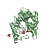
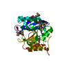
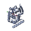
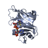
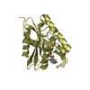
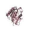
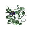
| Title | CIRCULARLY PERMUTED (1-3,1-4)-BETA-D-GLUCAN 4-GLUCANOHYDROLASE CPA16M-84 | ||||||
 Components Components | CIRCULARLY PERMUTED (1-3,1-4)-BETA-D-GLUCAN 4-GLUCANOHYDROLASE | ||||||
 Keywords Keywords | HYDROLASE / GLUCANASE / CIRCULAR PERMUTATION | ||||||
| Function / homology |  Function and homology information Function and homology information | ||||||
| Biological species |  Paenibacillus macerans (bacteria) Paenibacillus macerans (bacteria) | ||||||
| Method |  X-RAY DIFFRACTION / MOLECULAR REPLACEMENT / Resolution: 1.8 Å X-RAY DIFFRACTION / MOLECULAR REPLACEMENT / Resolution: 1.8 Å | ||||||
 Authors Authors | Ay, J. / Heinemann, U. | ||||||
 Citation Citation | Journal: Proteins / Year: 1998 Title: Crystal structures and properties of de novo circularly permuted 1,3-1,4-beta-glucanases. Authors: Ay, J. / Hahn, M. / Decanniere, K. / Piotukh, K. / Borriss, R. / Heinemann, U. | ||||||
| History |
|
- Structure visualization
| Structure viewer | Molecule: MolmilJmol/JSmol |
|---|
- Downloads & links
-Download
| PDBx/mmCIF format | 1ajk.cif.gz | 103.3 KB | Display | PDBx/mmCIF format |
|---|---|---|---|---|
| PDB format | pdb1ajk.ent.gz | 77.7 KB | Display | PDB format |
| PDBx/mmJSON format | 1ajk.json.gz | Tree view | PDBx/mmJSON format | |
| Others |  Other downloads Other downloads |
-Validation report
| Arichive directory | https://data.pdbj.org/pub/pdb/validation_reports/aj/1ajkftp://data.pdbj.org/pub/pdb/validation_reports/aj/1ajk | HTTPS FTP |
|---|
-Related structure data
| Related structure data |  1ajoC 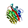 2ayhS S: Starting model for refinement C: citing same article ( |
|---|---|
| Similar structure data |


-Links
 PDBj
PDBj
- Assembly
| Deposited unit | 
| ||||||||
|---|---|---|---|---|---|---|---|---|---|
| 1 | 
| ||||||||
| 2 | 
| ||||||||
| Unit cell |
| ||||||||
| Noncrystallographic symmetry (NCS) | NCS oper: (Code: given Matrix: (-0.82105, -0.03541, -0.56976), Vector: |
-Components
| #1: Protein | Mass: 23935.221 Da / Num. of mol.: 2 Source method: isolated from a genetically manipulated source Source: (gene. exp.) Paenibacillus macerans (bacteria) / Cell line: DH5ALPHA / Plasmid: PTZ19R / Cell line (production host): DH5ALPHA / Production host: #2: Chemical |   Mass: 40.078 Da / Num. of mol.: 2 / Source method: obtained synthetically / Formula: Ca Mass: 40.078 Da / Num. of mol.: 2 / Source method: obtained synthetically / Formula: Ca#3: Chemical | ChemComp-PO4 / |   Mass: 94.971 Da / Num. of mol.: 1 / Source method: obtained synthetically / Formula: PO4 Mass: 94.971 Da / Num. of mol.: 1 / Source method: obtained synthetically / Formula: PO4#4: Chemical |   Mass: 238.305 Da / Num. of mol.: 2 / Source method: obtained synthetically / Formula: C8H18N2O4S / Comment: pH buffer*YM Mass: 238.305 Da / Num. of mol.: 2 / Source method: obtained synthetically / Formula: C8H18N2O4S / Comment: pH buffer*YM#5: Water | ChemComp-HOH / |  Mass: 18.015 Da / Num. of mol.: 303 / Source method: isolated from a natural source / Formula: H2O Mass: 18.015 Da / Num. of mol.: 303 / Source method: isolated from a natural source / Formula: H2OHas protein modification | Y | Nonpolymer details | BOTH INDEPENDENT MOLECULES OF CPA16M-84 SHOW A CATION BINDING SITE WITH A CALCIUM ION IN OCTAHEDRAL ...BOTH INDEPENDEN | |
|---|
-Experimental details
-Experiment
| Experiment | Method: X-RAY DIFFRACTION / Number of used crystals: 1 |
|---|
- Sample preparation
| Crystal | Density Matthews: 2.1 Å3/Da / Density % sol: 41 % Description: ONE LOW- AND ONE HIGH-RESOLUTION DATA SET WERE COLLECTED FROM ONE CRYSTAL. | ||||||||||||||||||||||||||||||||||||||||||||||||
|---|---|---|---|---|---|---|---|---|---|---|---|---|---|---|---|---|---|---|---|---|---|---|---|---|---|---|---|---|---|---|---|---|---|---|---|---|---|---|---|---|---|---|---|---|---|---|---|---|---|
| Crystal grow | Method: vapor diffusion, sitting drop / pH: 7 Details: SITTING DROP METHOD: A SOLUTION OF 12 MG OF PROTEIN PER ML IN 10 MM HEPES, PH 7.0, 2 MM CA-CHLORIDE, MIXED WITH AN EQUAL VOLUME OF 50 MM K-PHOSPHATE, PH 7.0 AND 20% (BY WEIGHT) PEG 8000, ...Details: SITTING DROP METHOD: A SOLUTION OF 12 MG OF PROTEIN PER ML IN 10 MM HEPES, PH 7.0, 2 MM CA-CHLORIDE, MIXED WITH AN EQUAL VOLUME OF 50 MM K-PHOSPHATE, PH 7.0 AND 20% (BY WEIGHT) PEG 8000, vapor diffusion - sitting drop | ||||||||||||||||||||||||||||||||||||||||||||||||
| Crystal | *PLUS | ||||||||||||||||||||||||||||||||||||||||||||||||
| Crystal grow | *PLUS Method: vapor diffusion, sitting drop | ||||||||||||||||||||||||||||||||||||||||||||||||
| Components of the solutions | *PLUS
|
-Data collection
| Diffraction | Mean temperature: 294 K |
|---|---|
| Diffraction source | Source: ROTATING ANODE / Type: RIGAKU FR-D / Wavelength: 1.5418 |
| Detector | Type: MARRESEARCH / Detector: IMAGE PLATE / Date: Apr 1, 1995 / Details: COLLIMATOR |
| Radiation | Monochromator: GRAPHITE(002) / Monochromatic (M) / Laue (L): M / Scattering type: x-ray |
| Radiation wavelength | Wavelength: 1.5418 Å / Relative weight: 1 |
| Reflection | Resolution: 1.76→33.33 Å / Num. obs: 38698 / % possible obs: 98.3 % / Redundancy: 3.4 % / Biso Wilson estimate: 19.34 Å2 / Rmerge(I) obs: 0.056 / Rsym value: 0.056 / Net I/σ(I): 5.8 |
| Reflection shell | Resolution: 1.76→1.83 Å / Redundancy: 3.2 % / Rmerge(I) obs: 0.164 / Mean I/σ(I) obs: 4.5 / Rsym value: 0.16 / % possible all: 94.4 |
| Reflection | *PLUS Num. measured all: 132154 |
| Reflection shell | *PLUS % possible obs: 94.4 % |
- Processing
| Software |
| ||||||||||||||||||||||||||||||||||||||||||||||||||||||||||||||||||||||||||||||||||||
|---|---|---|---|---|---|---|---|---|---|---|---|---|---|---|---|---|---|---|---|---|---|---|---|---|---|---|---|---|---|---|---|---|---|---|---|---|---|---|---|---|---|---|---|---|---|---|---|---|---|---|---|---|---|---|---|---|---|---|---|---|---|---|---|---|---|---|---|---|---|---|---|---|---|---|---|---|---|---|---|---|---|---|---|---|---|
| Refinement | Method to determine structure: MOLECULAR REPLACEMENT Starting model: PDB ENTRY 2AYH Resolution: 1.8→33.33 Å / Cross valid method: THROUGHOUT / σ(F): 0
| ||||||||||||||||||||||||||||||||||||||||||||||||||||||||||||||||||||||||||||||||||||
| Displacement parameters | Biso mean: 20.83 Å2 | ||||||||||||||||||||||||||||||||||||||||||||||||||||||||||||||||||||||||||||||||||||
| Refine analyze | Luzzati coordinate error obs: 0.3 Å / Luzzati d res low obs: 33.3 Å | ||||||||||||||||||||||||||||||||||||||||||||||||||||||||||||||||||||||||||||||||||||
| Refinement step | Cycle: LAST / Resolution: 1.8→33.33 Å
| ||||||||||||||||||||||||||||||||||||||||||||||||||||||||||||||||||||||||||||||||||||
| Refine LS restraints |
| ||||||||||||||||||||||||||||||||||||||||||||||||||||||||||||||||||||||||||||||||||||
| Software | *PLUS Name: REFMAC / Classification: refinement | ||||||||||||||||||||||||||||||||||||||||||||||||||||||||||||||||||||||||||||||||||||
| Refinement | *PLUS Num. reflection all: 35910 / Rfactor all: 0.168 | ||||||||||||||||||||||||||||||||||||||||||||||||||||||||||||||||||||||||||||||||||||
| Solvent computation | *PLUS | ||||||||||||||||||||||||||||||||||||||||||||||||||||||||||||||||||||||||||||||||||||
| Displacement parameters | *PLUS |
