 Movie
Movie Controller

[English] 日本語
 Yorodumi
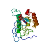
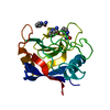
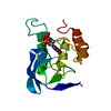
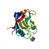
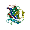
- PDB-1a3u: STAPHYLOCOCCAL NUCLEASE, CYCLOHEXANE THIOL DISULFIDE TO V23C VARIANT -
+ Open data

- Basic information
| Entry | Database: PDB / ID: 1a3u | ||||||
|---|---|---|---|---|---|---|---|
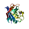
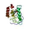
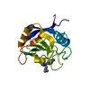
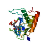
| Title | STAPHYLOCOCCAL NUCLEASE, CYCLOHEXANE THIOL DISULFIDE TO V23C VARIANT | ||||||
 Components Components | STAPHYLOCOCCAL NUCLEASE | ||||||
 Keywords Keywords | NUCLEASE / UNNATURAL AMINO ACID / HYDROLASE / ENDONUCLEASE | ||||||
| Function / homology |  Function and homology information Function and homology informationmicrococcal nuclease / 3' overhang single-stranded DNA endonuclease activity / nucleic acid binding / extracellular region / membrane / metal ion binding Similarity search - Function | ||||||
| Biological species |   Staphylococcus aureus (bacteria) Staphylococcus aureus (bacteria) | ||||||
| Method |  X-RAY DIFFRACTION / MOLECULAR REPLACEMENT / Resolution: 2.05 Å X-RAY DIFFRACTION / MOLECULAR REPLACEMENT / Resolution: 2.05 Å | ||||||
 Authors Authors | Wynn, R. / Harkins, P.C. / Richards, F.M. / Fox, R.O. | ||||||
 Citation Citation | Journal: Protein Sci. / Year: 1997 Title: Comparison of straight chain and cyclic unnatural amino acids embedded in the core of staphylococcal nuclease. Authors: Wynn, R. / Harkins, P.C. / Richards, F.M. / Fox, R.O. #1: Journal: Protein Sci. / Year: 1996Title: Mobile Unnatural Amino Acid Side Chains in the Core of Staphylococcal Nuclease Authors: Wynn, R. / Harkins, P.C. / Richards, F.M. / Fox, R.O. #2: Journal: Protein Sci. / Year: 1995Title: Interactions in Nonnative and Truncated Forms of Staphylococcal Nuclease as Indicated by Mutational Free Energy Changes Authors: Wynn, R. / Anderson, C.L. / Richards, F.M. / Fox, R.O. | ||||||
| History |
|
- Structure visualization
| Structure viewer | Molecule: MolmilJmol/JSmol |
|---|
- Downloads & links
-Download
| PDBx/mmCIF format | 1a3u.cif.gz | 45.4 KB | Display | PDBx/mmCIF format |
|---|---|---|---|---|
| PDB format | pdb1a3u.ent.gz | 34.3 KB | Display | PDB format |
| PDBx/mmJSON format | 1a3u.json.gz | Tree view | PDBx/mmJSON format | |
| Others |  Other downloads Other downloads |
-Validation report
| Arichive directory | https://data.pdbj.org/pub/pdb/validation_reports/a3/1a3uftp://data.pdbj.org/pub/pdb/validation_reports/a3/1a3u | HTTPS FTP |
|---|
-Related structure data
| Related structure data | 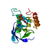 1a3tC 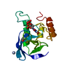 1a3vC 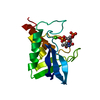 1sncS S: Starting model for refinement C: citing same article ( |
|---|---|
| Similar structure data |

-Links
 PDBj
PDBj- Assembly
| Deposited unit | 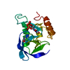
| ||||||||
|---|---|---|---|---|---|---|---|---|---|
| 1 |
| ||||||||
| Unit cell |
|
-Components
| #1: Protein | Mass: 16961.551 Da / Num. of mol.: 1 / Mutation: V23C, CYCLOHEXANE THIOL DISULFIDE Source method: isolated from a genetically manipulated source Details: VARIANT FORMED BY CHEMICAL MODIFICATION OF SOLE CYSTEINE RESIDUE, PDTP, 3',5'-THYMIDINE DIPHOSPHATE Source: (gene. exp.) Staphylococcus aureus (bacteria) / Production host: |
|---|---|
| #2: Chemical | ChemComp-CA /   Mass: 40.078 Da / Num. of mol.: 1 / Source method: obtained synthetically / Formula: Ca Mass: 40.078 Da / Num. of mol.: 1 / Source method: obtained synthetically / Formula: Ca |
| #3: Chemical | ChemComp-THP /   Type: DNA linking / Mass: 402.188 Da / Num. of mol.: 1 / Source method: obtained synthetically / Formula: C10H16N2O11P2 Type: DNA linking / Mass: 402.188 Da / Num. of mol.: 1 / Source method: obtained synthetically / Formula: C10H16N2O11P2 |
| #4: Water | ChemComp-HOH /  Mass: 18.015 Da / Num. of mol.: 41 / Source method: isolated from a natural source / Formula: H2O Mass: 18.015 Da / Num. of mol.: 41 / Source method: isolated from a natural source / Formula: H2O |
-Experimental details
-Experiment
| Experiment | Method: X-RAY DIFFRACTION / Number of used crystals: 1 |
|---|
- Sample preparation
| Crystal | Density Matthews: 2.17 Å3/Da / Density % sol: 43.36 % | ||||||||||||||||||||||||||||||||||||||||||||||||
|---|---|---|---|---|---|---|---|---|---|---|---|---|---|---|---|---|---|---|---|---|---|---|---|---|---|---|---|---|---|---|---|---|---|---|---|---|---|---|---|---|---|---|---|---|---|---|---|---|---|
| Crystal grow | Temperature: 277 K / pH: 8.15 Details: 10 MM POTASSIUM PHOSPHATE, PH 8.15 PROTEIN CONCENTRATION = 2 MGS/ML MPD =21% T=4 DEGREES C, temperature 277K | ||||||||||||||||||||||||||||||||||||||||||||||||
| Crystal grow | *PLUS Temperature: 4 ℃ / Method: vapor diffusionDetails: Loll, P.J., (1989) Proteins Struct. Funct. Genet., 5, 183. | ||||||||||||||||||||||||||||||||||||||||||||||||
| Components of the solutions | *PLUS
|
-Data collection
| Diffraction | Mean temperature: 278.15 K |
|---|---|
| Diffraction source | Source: ROTATING ANODE / Type: RIGAKU RUH2R / Wavelength: 1.5418 |
| Detector | Type: MACSCIENCE / Detector: IMAGE PLATE / Date: Jan 1, 1996 / Details: COLLIMATOR |
| Radiation | Monochromator: NI FILTER / Monochromatic (M) / Laue (L): M / Scattering type: x-ray |
| Radiation wavelength | Wavelength: 1.5418 Å / Relative weight: 1 |
| Reflection | Resolution: 2.05→6 Å / Num. obs: 7657 / % possible obs: 86.9 % / Observed criterion σ(I): 2 / Redundancy: 6.2 % / Rmerge(I) obs: 0.059 |
| Reflection shell | Resolution: 2.05→2.15 Å / % possible all: 72.4 |
| Reflection shell | *PLUS % possible obs: 72.4 % |
- Processing
| Software |
| ||||||||||||||||||
|---|---|---|---|---|---|---|---|---|---|---|---|---|---|---|---|---|---|---|---|
| Refinement | Method to determine structure: MOLECULAR REPLACEMENT Starting model: PDB ENTRY 1SNC Resolution: 2.05→6 Å / σ(F): 2 /
| ||||||||||||||||||
| Refinement step | Cycle: LAST / Resolution: 2.05→6 Å
| ||||||||||||||||||
| Xplor file |
|
