 Movie
Movie Controller

[English] 日本語
 Yorodumi
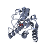
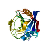
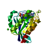
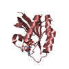
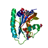
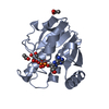
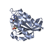
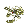
- PDB-1a33: PEPTIDYLPROLYL ISOMERASE, CYCLOPHILIN-LIKE DOMAIN FROM BRUGIA MALAYI -
+ Open data

- Basic information
| Entry | Database: PDB / ID: 1a33 | ||||||
|---|---|---|---|---|---|---|---|
| Title | PEPTIDYLPROLYL ISOMERASE, CYCLOPHILIN-LIKE DOMAIN FROM BRUGIA MALAYI | ||||||
 Components Components | PEPTIDYLPROLYL ISOMERASE | ||||||
 Keywords Keywords | ISOMERASE / PEPTIDYL-PROLYL CIS-TRANS / PEPTIDYLPROLYL ISOMERASE | ||||||
| Function / homology |  Function and homology information Function and homology informationcyclosporin A binding / peptidylprolyl isomerase / peptidyl-prolyl cis-trans isomerase activity / protein folding / mitochondrion Similarity search - Function | ||||||
| Biological species |  Brugia malayi (agent of lymphatic filariasis) Brugia malayi (agent of lymphatic filariasis) | ||||||
| Method |  X-RAY DIFFRACTION / MOLECULAR REPLACEMENT / Resolution: 2.15 Å X-RAY DIFFRACTION / MOLECULAR REPLACEMENT / Resolution: 2.15 Å | ||||||
 Authors Authors | Mikol, V. / Ma, D. / Carlow, C.K.S. | ||||||
 Citation Citation | Journal: Protein Sci. / Year: 1998 Title: Crystal structure of the cyclophilin-like domain from the parasitic nematode Brugia malayi. Authors: Mikol, V. / Ma, D. / Carlow, C.K. | ||||||
| History |
|
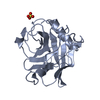
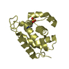
- Structure visualization
| Structure viewer | Molecule: MolmilJmol/JSmol |
|---|
- Downloads & links
-Download
| PDBx/mmCIF format | 1a33.cif.gz | 49.5 KB | Display | PDBx/mmCIF format |
|---|---|---|---|---|
| PDB format | pdb1a33.ent.gz | 35.3 KB | Display | PDB format |
| PDBx/mmJSON format | 1a33.json.gz | Tree view | PDBx/mmJSON format | |
| Others |  Other downloads Other downloads |
-Validation report
| Summary document | 1a33_validation.pdf.gz | 406.7 KB | Display | wwPDB validaton report |
|---|---|---|---|---|
| Full document | 1a33_full_validation.pdf.gz | 408 KB | Display | |
| Data in XML | 1a33_validation.xml.gz | 10.9 KB | Display | |
| Data in CIF | 1a33_validation.cif.gz | 16 KB | Display | |
| Arichive directory | https://data.pdbj.org/pub/pdb/validation_reports/a3/1a33ftp://data.pdbj.org/pub/pdb/validation_reports/a3/1a33 | HTTPS FTP |
-Related structure data
| Related structure data | 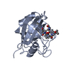 1cwaS S: Starting model for refinement |
|---|---|
| Similar structure data |
-Links
 PDBj
PDBj

- Assembly
| Deposited unit | 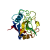
| ||||||||
|---|---|---|---|---|---|---|---|---|---|
| 1 |
| ||||||||
| Unit cell |
|
-Components
| #1: Protein | Mass: 19504.418 Da / Num. of mol.: 1 / Fragment: CYCLOPHILIN-LIKE DOMAIN Source method: isolated from a genetically manipulated source Source: (gene. exp.) Brugia malayi (agent of lymphatic filariasis)Gene: BMCYP-1 / Plasmid: PMAL-C2 / Gene (production host): MBP FUSION PROTEIN / Production host:  |
|---|---|
| #2: Water | ChemComp-HOH /  Mass: 18.015 Da / Num. of mol.: 234 / Source method: isolated from a natural source / Formula: H2O Mass: 18.015 Da / Num. of mol.: 234 / Source method: isolated from a natural source / Formula: H2O |
-Experimental details
-Experiment
| Experiment | Method: X-RAY DIFFRACTION / Number of used crystals: 1 |
|---|
- Sample preparation
| Crystal | Density Matthews: 2.67 Å3/Da / Density % sol: 54.1 % | ||||||||||||||||||||||||||||||
|---|---|---|---|---|---|---|---|---|---|---|---|---|---|---|---|---|---|---|---|---|---|---|---|---|---|---|---|---|---|---|---|
| Crystal grow | pH: 6 Details: THE INITIAL 6 UL DROP CONSISTED OF 50 MM MES-NAOH PH 6.0, 0.9 M (NH4)2SO4, 1MM PROTEIN. THE 1 ML RESERVOIR SOLUTION CONSISTED OF 100 MM MES-NAOH PH 6.0, 1.8 M (NH4)2SO4 | ||||||||||||||||||||||||||||||
| Crystal grow | *PLUS Temperature: 19 ℃ / Method: vapor diffusion, hanging drop | ||||||||||||||||||||||||||||||
| Components of the solutions | *PLUS
|
-Data collection
| Diffraction | Mean temperature: 95 K |
|---|---|
| Diffraction source | Source: ROTATING ANODE / Type: ENRAF-NONIUS FR591 / Wavelength: 1.5418 |
| Detector | Type: MACSCIENCE / Detector: IMAGE PLATE / Date: Jul 1, 1996 / Details: MIRRORS |
| Radiation | Monochromator: NI FILTER / Monochromatic (M) / Laue (L): M / Scattering type: x-ray |
| Radiation wavelength | Wavelength: 1.5418 Å / Relative weight: 1 |
| Reflection | Resolution: 2.15→10 Å / Num. obs: 13361 / % possible obs: 98.6 % / Observed criterion σ(I): 0 / Redundancy: 6.4 % / Rmerge(I) obs: 0.05 / Rsym value: 0.05 / Net I/σ(I): 36 |
| Reflection shell | Resolution: 2.15→2.23 Å / Redundancy: 6 % / Rmerge(I) obs: 0.08 / Mean I/σ(I) obs: 22 / Rsym value: 0.08 / % possible all: 96 |
| Reflection | *PLUS Num. measured all: 84704 |
| Reflection shell | *PLUS % possible obs: 98.6 % |
- Processing
| Software |
| ||||||||||||||||||||||||||||||||||||||||||||||||||||||||||||
|---|---|---|---|---|---|---|---|---|---|---|---|---|---|---|---|---|---|---|---|---|---|---|---|---|---|---|---|---|---|---|---|---|---|---|---|---|---|---|---|---|---|---|---|---|---|---|---|---|---|---|---|---|---|---|---|---|---|---|---|---|---|
| Refinement | Method to determine structure: MOLECULAR REPLACEMENT Starting model: PDB ENTRY 1CWA Resolution: 2.15→8 Å / Data cutoff high absF: 10000000 / Data cutoff low absF: 0 / Cross valid method: THROUGOUT / σ(F): 0
| ||||||||||||||||||||||||||||||||||||||||||||||||||||||||||||
| Displacement parameters | Biso mean: 12 Å2 | ||||||||||||||||||||||||||||||||||||||||||||||||||||||||||||
| Refinement step | Cycle: LAST / Resolution: 2.15→8 Å
| ||||||||||||||||||||||||||||||||||||||||||||||||||||||||||||
| Refine LS restraints |
| ||||||||||||||||||||||||||||||||||||||||||||||||||||||||||||
| LS refinement shell | Resolution: 2.15→2.25 Å / Rfactor Rfree error: 0 / Total num. of bins used: 8
| ||||||||||||||||||||||||||||||||||||||||||||||||||||||||||||
| Xplor file |
| ||||||||||||||||||||||||||||||||||||||||||||||||||||||||||||
| Software | *PLUS Name: X-PLOR / Version: 3.851 / Classification: refinement | ||||||||||||||||||||||||||||||||||||||||||||||||||||||||||||
| Refine LS restraints | *PLUS
|
