 Movie
Movie Controller

[English] 日本語
 Yorodumi
- PDB-1a2o: STRUCTURAL BASIS FOR METHYLESTERASE CHEB REGULATION BY A PHOSPHOR... -
+ Open data

- Basic information
| Entry | Database: PDB / ID: 1a2o | ||||||
|---|---|---|---|---|---|---|---|
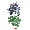
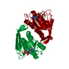
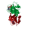
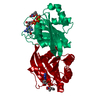
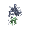
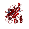
| Title | STRUCTURAL BASIS FOR METHYLESTERASE CHEB REGULATION BY A PHOSPHORYLATION-ACTIVATED DOMAIN | ||||||
 Components Components | CHEB METHYLESTERASE | ||||||
 Keywords Keywords | BACTERIAL CHEMOTAXIS / ADAPTATION / SERINE HYDROLASE | ||||||
| Function / homology |  Function and homology information Function and homology informationprotein-glutamate methylesterase / protein-glutamate methylesterase activity / protein-glutamine glutaminase activity / protein-glutamine glutaminase / phosphorelay response regulator activity / chemotaxis / signal transduction / cytoplasm Similarity search - Function | ||||||
| Biological species |  Salmonella typhimurium (bacteria) Salmonella typhimurium (bacteria) | ||||||
| Method |  X-RAY DIFFRACTION / MOLECULAR REPLACEMENT / Resolution: 2.4 Å X-RAY DIFFRACTION / MOLECULAR REPLACEMENT / Resolution: 2.4 Å | ||||||
 Authors Authors | Djordjevic, S. / Goudreau, P.N. / Xu, Q. / Stock, A.M. / West, A.H. | ||||||
 Citation Citation | Journal: Proc.Natl.Acad.Sci.USA / Year: 1998 Title: Structural basis for methylesterase CheB regulation by a phosphorylation-activated domain. Authors: Djordjevic, S. / Goudreau, P.N. / Xu, Q. / Stock, A.M. / West, A.H. | ||||||
| History |
|
- Structure visualization
| Structure viewer | Molecule: MolmilJmol/JSmol |
|---|
- Downloads & links
-Download
| PDBx/mmCIF format | 1a2o.cif.gz | 147.1 KB | Display | PDBx/mmCIF format |
|---|---|---|---|---|
| PDB format | pdb1a2o.ent.gz | 116.8 KB | Display | PDB format |
| PDBx/mmJSON format | 1a2o.json.gz | Tree view | PDBx/mmJSON format | |
| Others |  Other downloads Other downloads |
-Validation report
| Arichive directory | https://data.pdbj.org/pub/pdb/validation_reports/a2/1a2oftp://data.pdbj.org/pub/pdb/validation_reports/a2/1a2o | HTTPS FTP |
|---|
-Related structure data
| Related structure data | 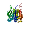 1chdS S: Starting model for refinement |
|---|---|
| Similar structure data |


-Links
 PDBj
PDBj


- Assembly
| Deposited unit | 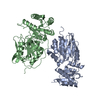
| ||||||||||||
|---|---|---|---|---|---|---|---|---|---|---|---|---|---|
| 1 | 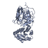
| ||||||||||||
| 2 | 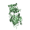
| ||||||||||||
| Unit cell |
| ||||||||||||
| Noncrystallographic symmetry (NCS) | NCS oper:
|
-Components
| #1: Protein | Mass: 37598.852 Da / Num. of mol.: 2 Source method: isolated from a genetically manipulated source Source: (gene. exp.) Salmonella typhimurium (bacteria) / Cellular location: CYTOPLASM / Plasmid: PME30 / Cell (production host): HB101 / Cellular location (production host): CYTOPLASM / Production host: References: UniProt: P04042, protein-glutamate methylesterase #2: Water | ChemComp-HOH / |  Mass: 18.015 Da / Num. of mol.: 411 / Source method: isolated from a natural source / Formula: H2O Mass: 18.015 Da / Num. of mol.: 411 / Source method: isolated from a natural source / Formula: H2O |
|---|
-Experimental details
-Experiment
| Experiment | Method: X-RAY DIFFRACTION / Number of used crystals: 1 |
|---|
- Sample preparation
| Crystal | Density Matthews: 2.87 Å3/Da / Density % sol: 57.17 % | ||||||||||||||||||||||||||||||||||||||||||||||||||||||||
|---|---|---|---|---|---|---|---|---|---|---|---|---|---|---|---|---|---|---|---|---|---|---|---|---|---|---|---|---|---|---|---|---|---|---|---|---|---|---|---|---|---|---|---|---|---|---|---|---|---|---|---|---|---|---|---|---|---|
| Crystal grow | pH: 6.5 / Details: pH 6.5 | ||||||||||||||||||||||||||||||||||||||||||||||||||||||||
| Crystal grow | *PLUS Method: vapor diffusion, hanging drop / Details: used to seeding | ||||||||||||||||||||||||||||||||||||||||||||||||||||||||
| Components of the solutions | *PLUS
|
-Data collection
| Diffraction | Mean temperature: 100 K |
|---|---|
| Diffraction source | Source: ROTATING ANODE / Type: RIGAKU RUH2R / Wavelength: 1.5418 |
| Detector | Type: RIGAKU / Detector: IMAGE PLATE / Date: Jan 1, 1997 / Details: MIRRORS |
| Radiation | Monochromator: NI FILTER / Monochromatic (M) / Laue (L): M / Scattering type: x-ray |
| Radiation wavelength | Wavelength: 1.5418 Å / Relative weight: 1 |
| Reflection | Resolution: 2.4→15 Å / Num. obs: 32281 / % possible obs: 96.9 % / Observed criterion σ(I): 2 / Redundancy: 3 % / Biso Wilson estimate: 38.4 Å2 / Rsym value: 0.054 / Net I/σ(I): 7.5 |
| Reflection shell | Resolution: 2.4→2.46 Å / Redundancy: 3.1 % / Mean I/σ(I) obs: 4.1 / Rsym value: 0.163 / % possible all: 98.5 |
| Reflection | *PLUS Num. measured all: 97667 / Rmerge(I) obs: 0.054 |
- Processing
| Software |
| ||||||||||||||||||||||||||||||||||||||||||||||||||||||||||||||||||||||||||||||||||||
|---|---|---|---|---|---|---|---|---|---|---|---|---|---|---|---|---|---|---|---|---|---|---|---|---|---|---|---|---|---|---|---|---|---|---|---|---|---|---|---|---|---|---|---|---|---|---|---|---|---|---|---|---|---|---|---|---|---|---|---|---|---|---|---|---|---|---|---|---|---|---|---|---|---|---|---|---|---|---|---|---|---|---|---|---|---|
| Refinement | Method to determine structure: MOLECULAR REPLACEMENT Starting model: PDB ENTRY 1CHD Resolution: 2.4→10 Å / Cross valid method: FREE R / σ(F): 0
| ||||||||||||||||||||||||||||||||||||||||||||||||||||||||||||||||||||||||||||||||||||
| Displacement parameters | Biso mean: 23.7 Å2 | ||||||||||||||||||||||||||||||||||||||||||||||||||||||||||||||||||||||||||||||||||||
| Refinement step | Cycle: LAST / Resolution: 2.4→10 Å
| ||||||||||||||||||||||||||||||||||||||||||||||||||||||||||||||||||||||||||||||||||||
| Refine LS restraints |
| ||||||||||||||||||||||||||||||||||||||||||||||||||||||||||||||||||||||||||||||||||||
| Software | *PLUS Name: REFMAC / Classification: refinement | ||||||||||||||||||||||||||||||||||||||||||||||||||||||||||||||||||||||||||||||||||||
| Refinement | *PLUS Num. reflection all: 32281 / Rfactor all: 0.222 / Rfactor Rfree: 0.288 | ||||||||||||||||||||||||||||||||||||||||||||||||||||||||||||||||||||||||||||||||||||
| Solvent computation | *PLUS | ||||||||||||||||||||||||||||||||||||||||||||||||||||||||||||||||||||||||||||||||||||
| Displacement parameters | *PLUS |
