 Movie
Movie Controller

[English] 日本語
 Yorodumi
- PDB-13eu: Cryo-EM structure of HAdV-C6 hexon trimer in complex with human c... -
+ Open data

- Basic information
| Entry | Database: PDB / ID: 13eu | ||||||||||||||||||||||||
|---|---|---|---|---|---|---|---|---|---|---|---|---|---|---|---|---|---|---|---|---|---|---|---|---|---|
| Title | Cryo-EM structure of HAdV-C6 hexon trimer in complex with human coagulation factor X (FX) | ||||||||||||||||||||||||
 Components Components |
| ||||||||||||||||||||||||
 Keywords Keywords | VIRAL PROTEIN / adenovirus / hexon / coagulation factor X / virus-host interaction / cryo-EM / virus capsid | ||||||||||||||||||||||||
| Function / homology |  Function and homology information Function and homology informationT=25 icosahedral viral capsid / coagulation factor Xa / microtubule-dependent intracellular transport of viral material towards nucleus / Defective factor IX causes thrombophilia / Defective cofactor function of FVIIIa variant / Defective F9 variant does not activate FX / Transport of gamma-carboxylated protein precursors from the endoplasmic reticulum to the Golgi apparatus / Gamma-carboxylation of protein precursors / Removal of aminoterminal propeptides from gamma-carboxylated proteins / Initiation of coagulation cascade ...T=25 icosahedral viral capsid / coagulation factor Xa / microtubule-dependent intracellular transport of viral material towards nucleus / Defective factor IX causes thrombophilia / Defective cofactor function of FVIIIa variant / Defective F9 variant does not activate FX / Transport of gamma-carboxylated protein precursors from the endoplasmic reticulum to the Golgi apparatus / Gamma-carboxylation of protein precursors / Removal of aminoterminal propeptides from gamma-carboxylated proteins / Initiation of coagulation cascade / Amplification and propagation of coagulation cascade / positive regulation of TOR signaling / Regulation of clotting cascade / blood coagulation / phospholipid binding / Golgi lumen / host cell / positive regulation of cell migration / endoplasmic reticulum lumen / serine-type endopeptidase activity / external side of plasma membrane / calcium ion binding / symbiont entry into host cell / host cell nucleus / structural molecule activity / proteolysis / extracellular region / plasma membrane Similarity search - Function | ||||||||||||||||||||||||
| Biological species |  Human adenovirus 6 Human adenovirus 6 Homo sapiens (human) Homo sapiens (human) | ||||||||||||||||||||||||
| Method | ELECTRON MICROSCOPY / single particle reconstruction / cryo EM / Resolution: 3.26 Å | ||||||||||||||||||||||||
 Authors Authors | Ma, O.X. / Reddy, V.S. | ||||||||||||||||||||||||
| Funding support |  United States, 1items United States, 1items
| ||||||||||||||||||||||||
 Citation Citation | Journal: PLoS Pathog / Year: 2026 Title: Structural requirements of blood factors binding to soluble hexon trimers with implications for adenovirus cell targeting and immune evasion. Authors: Olivia X Ma / Shao-Chia Lu / Haley E Mudrick / Mary E Barry / Jarrod B French / Michael A Barry / Vijay S Reddy / Abstract: Human adenovirus serotype 5 (HAdV-C5) is widely used as a gene delivery vector in both experimental and clinical settings. Upon intravenous administration, HAdV-C5 exhibits strong liver tropism, ...Human adenovirus serotype 5 (HAdV-C5) is widely used as a gene delivery vector in both experimental and clinical settings. Upon intravenous administration, HAdV-C5 exhibits strong liver tropism, largely mediated by interactions between its major capsid protein, hexon (Hx), and coagulation factor X (FX). In contrast, the closely related species C adenovirus 6 (HAdV-C6) also targets the liver but shows reduced dependency on coagulation factors, whereas species D adenovirus 26 (HAdV-D26) does not bind coagulation factors altogether. To define the structural basis of this serotype-specific host factor recognition, we determined high-resolution cryo-electron microscopy structures of isolated hexon trimers from HAdV-C5 and HAdV-C6 in complex with coagulation factors FX and prothrombin (factor II, FII). The resulting atomic models reveal conserved binding interfaces involving the γ-carboxyglutamic acid (Gla) domains of both coagulation factors and the hypervariable regions HVR5 and HVR7 lining the surface cavities of HAdV-C5 and HAdV-C6 hexons. Structures of hexon complexes formed by co-incubation with both FX and FII further reveal serotype-specific binding preferences under physiologically relevant conditions, showing that HAdV-C5 hexon preferentially engages FX, whereas HAdV-C6 hexon favors FII. By contrast, HAdV-D26 hexon does not bind either factor, likely due to an insertion constrained by proline residues in the HVR5 loop that restricts the factor access to the hexon cavity. Together, these findings provide a detailed structural framework for adenovirus-coagulation factor interactions and support the rational engineering of adenovirus vectors with improved targeting and safety profiles. | ||||||||||||||||||||||||
| History |
|
- Structure visualization
| Structure viewer | Molecule: MolmilJmol/JSmol |
|---|
- Downloads & links
-Download
| PDBx/mmCIF format | 13eu.cif.gz | 572.1 KB | Display | PDBx/mmCIF format |
|---|---|---|---|---|
| PDB format | pdb13eu.ent.gz | 457 KB | Display | PDB format |
| PDBx/mmJSON format | 13eu.json.gz | Tree view | PDBx/mmJSON format | |
| Others |  Other downloads Other downloads |
-Validation report
| Arichive directory | https://data.pdbj.org/pub/pdb/validation_reports/3e/13euftp://data.pdbj.org/pub/pdb/validation_reports/3e/13eu | HTTPS FTP |
|---|
-Related structure data
| Related structure data |  77027MC  13cmC  13djC  13dmC  13erC  13esC M: map data used to model this data C: citing same article ( |
|---|---|
| Similar structure data |


-Links
 PDBj
PDBj

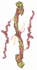
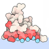
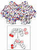

- Assembly
| Deposited unit | 
|
|---|---|
| 1 |
|
-Components
| #1: Protein | Mass: 108635.133 Da / Num. of mol.: 3 Source method: isolated from a genetically manipulated source Details: The modeled structure lacks the N-terminal residues 1-6, an internal region corresponding to residues 139-164, and a short segment around residues 445-452, as well as the C-terminal residues ...Details: The modeled structure lacks the N-terminal residues 1-6, an internal region corresponding to residues 139-164, and a short segment around residues 445-452, as well as the C-terminal residues 948-952. These regions were not included in the model due to the absence of well-defined cryo-EM density. Source: (gene. exp.) Human adenovirus 6 / Gene: Hexon, L3 / Production host: Human adenovirus 6 / References: UniProt: B2ZWX4#2: Protein | | Mass: 47560.695 Da / Num. of mol.: 1 / Source method: isolated from a natural source Details: The modeled coagulation factor X (FX) corresponds to the N-terminal Gla domain, while the signal peptide, propeptide, and the remainder of the protein (including EGF-like domains and the ...Details: The modeled coagulation factor X (FX) corresponds to the N-terminal Gla domain, while the signal peptide, propeptide, and the remainder of the protein (including EGF-like domains and the protease domain) are not included in the model due to absence of interpretable cryo-EM density. The retained region contains multiple gamma-carboxyglutamic acid (Gla) residues required for calcium coordination. Source: (natural) Homo sapiens (human) / Tissue: blood / References: UniProt: P00742, coagulation factor Xa#3: Chemical | ChemComp-CA /   Mass: 40.078 Da / Num. of mol.: 7 / Source method: obtained synthetically / Formula: Ca / Feature type: SUBJECT OF INVESTIGATION Mass: 40.078 Da / Num. of mol.: 7 / Source method: obtained synthetically / Formula: Ca / Feature type: SUBJECT OF INVESTIGATIONHas ligand of interest | Y | Has protein modification | Y | |
|---|
-Experimental details
-Experiment
| Experiment | Method: ELECTRON MICROSCOPY |
|---|---|
| EM experiment | Aggregation state: PARTICLE / 3D reconstruction method: single particle reconstruction |
- Sample preparation
| Component | Name: Purified Human adenovirus type 6 hexon trimer in complex with FX Type: COMPLEX / Entity ID: #1-#2 / Source: NATURAL |
|---|---|
| Molecular weight | Value: 0.39 MDa / Experimental value: NO |
| Source (natural) | Organism: Human adenovirus 6 |
| Buffer solution | pH: 7.2 |
| Specimen | Embedding applied: NO / Shadowing applied: NO / Staining applied: NO / Vitrification applied: YES |
| Specimen support | Grid material: COPPER / Grid mesh size: 300 divisions/in. / Grid type: Quantifoil R1.2/1.3 |
| Vitrification | Instrument: FEI VITROBOT MARK IV / Cryogen name: ETHANE / Humidity: 98 % / Chamber temperature: 277 K |
- Electron microscopy imaging
| Experimental equipment |  Model: Titan Krios / Image courtesy: FEI Company |
|---|---|
| Microscopy | Model: TFS KRIOS |
| Electron gun | Electron source:  FIELD EMISSION GUN / Accelerating voltage: 300 kV / Illumination mode: FLOOD BEAM FIELD EMISSION GUN / Accelerating voltage: 300 kV / Illumination mode: FLOOD BEAM |
| Electron lens | Mode: BRIGHT FIELD / Nominal defocus max: 2400 nm / Nominal defocus min: 800 nm / Cs: 2.7 mm / Alignment procedure: COMA FREE |
| Specimen holder | Cryogen: NITROGEN / Specimen holder model: FEI TITAN KRIOS AUTOGRID HOLDER |
| Image recording | Electron dose: 51 e/Å2 / Film or detector model: GATAN K3 BIOCONTINUUM (6k x 4k) / Num. of real images: 3510 |
| EM imaging optics | Energyfilter slit width: 20 eV |
- Processing
| EM software |
| ||||||||||||||||||||||||
|---|---|---|---|---|---|---|---|---|---|---|---|---|---|---|---|---|---|---|---|---|---|---|---|---|---|
| CTF correction | Type: PHASE FLIPPING ONLY | ||||||||||||||||||||||||
| Particle selection | Num. of particles selected: 280503 | ||||||||||||||||||||||||
| 3D reconstruction | Resolution: 3.26 Å / Resolution method: FSC 0.143 CUT-OFF / Num. of particles: 68552 / Symmetry type: POINT | ||||||||||||||||||||||||
| Refinement | Cross valid method: NONE Stereochemistry target values: REAL-SPACE (WEIGHTED MAP SUM AT ATOM CENTERS) | ||||||||||||||||||||||||
| Refine LS restraints |
|
