 Movie
Movie Controller

[English] 日本語
 Yorodumi
- EMDB-77025: Cryo-EM structure of HAdV-C5 hexon trimer in complex with human c... -
+ Open data

- Basic information
| Entry |  | |||||||||
|---|---|---|---|---|---|---|---|---|---|---|
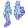
| Title | Cryo-EM structure of HAdV-C5 hexon trimer in complex with human coagulation factor X (FX) | |||||||||
 Map data Map data | EM map of the purified HAdV-C5 hexon trimer in complex with the coagulation factor X | |||||||||
 Sample Sample |
| |||||||||
 Keywords Keywords | adenovirus / HAdV-C5 / hexon / coagulation factor X / virus-host interaction / cryo-EM / virus capsid / VIRAL PROTEIN | |||||||||
| Function / homology |  Function and homology information Function and homology informationT=25 icosahedral viral capsid / coagulation factor Xa / Defective factor IX causes thrombophilia / Defective cofactor function of FVIIIa variant / Defective F9 variant does not activate FX / microtubule-dependent intracellular transport of viral material towards nucleus / : / positive regulation of TOR signaling / Transport of gamma-carboxylated protein precursors from the endoplasmic reticulum to the Golgi apparatus / : ...T=25 icosahedral viral capsid / coagulation factor Xa / Defective factor IX causes thrombophilia / Defective cofactor function of FVIIIa variant / Defective F9 variant does not activate FX / microtubule-dependent intracellular transport of viral material towards nucleus / : / positive regulation of TOR signaling / Transport of gamma-carboxylated protein precursors from the endoplasmic reticulum to the Golgi apparatus / : / Gamma-carboxylation of protein precursors / Removal of aminoterminal propeptides from gamma-carboxylated proteins / : / phospholipid binding / Golgi lumen / blood coagulation / viral capsid / host cell / positive regulation of cell migration / endoplasmic reticulum lumen / serine-type endopeptidase activity / external side of plasma membrane / calcium ion binding / symbiont entry into host cell / host cell nucleus / structural molecule activity / proteolysis / : / extracellular region / plasma membrane Similarity search - Function | |||||||||
| Biological species |  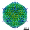 Human adenovirus 5 / Human adenovirus 5 /  Homo sapiens (human) Homo sapiens (human) | |||||||||
| Method | single particle reconstruction / cryo EM / Resolution: 3.23 Å | |||||||||
 Authors Authors | Ma OX / Reddy VS | |||||||||
| Funding support |  United States, 1 items United States, 1 items
| |||||||||
 Citation Citation | Journal: PLoS Pathog / Year: 2026 Title: Structural requirements of blood factors binding to soluble hexon trimers with implications for adenovirus cell targeting and immune evasion. Authors: Olivia X Ma / Shao-Chia Lu / Haley E Mudrick / Mary E Barry / Jarrod B French / Michael A Barry / Vijay S Reddy / Abstract: Human adenovirus serotype 5 (HAdV-C5) is widely used as a gene delivery vector in both experimental and clinical settings. Upon intravenous administration, HAdV-C5 exhibits strong liver tropism, ...Human adenovirus serotype 5 (HAdV-C5) is widely used as a gene delivery vector in both experimental and clinical settings. Upon intravenous administration, HAdV-C5 exhibits strong liver tropism, largely mediated by interactions between its major capsid protein, hexon (Hx), and coagulation factor X (FX). In contrast, the closely related species C adenovirus 6 (HAdV-C6) also targets the liver but shows reduced dependency on coagulation factors, whereas species D adenovirus 26 (HAdV-D26) does not bind coagulation factors altogether. To define the structural basis of this serotype-specific host factor recognition, we determined high-resolution cryo-electron microscopy structures of isolated hexon trimers from HAdV-C5 and HAdV-C6 in complex with coagulation factors FX and prothrombin (factor II, FII). The resulting atomic models reveal conserved binding interfaces involving the γ-carboxyglutamic acid (Gla) domains of both coagulation factors and the hypervariable regions HVR5 and HVR7 lining the surface cavities of HAdV-C5 and HAdV-C6 hexons. Structures of hexon complexes formed by co-incubation with both FX and FII further reveal serotype-specific binding preferences under physiologically relevant conditions, showing that HAdV-C5 hexon preferentially engages FX, whereas HAdV-C6 hexon favors FII. By contrast, HAdV-D26 hexon does not bind either factor, likely due to an insertion constrained by proline residues in the HVR5 loop that restricts the factor access to the hexon cavity. Together, these findings provide a detailed structural framework for adenovirus-coagulation factor interactions and support the rational engineering of adenovirus vectors with improved targeting and safety profiles. | |||||||||
| History |
|
- Structure visualization
| Supplemental images |
|---|

- Downloads & links
-EMDB archive
| Map data | emd_77025.map.gz | 55.8 MB | EMDB map data format | |
|---|---|---|---|---|
| Header (meta data) | emd-77025-v30.xmlemd-77025.xml | 23.8 KB 23.8 KB | Display Display | EMDB header |
| FSC (resolution estimation) | emd_77025_fsc.xml | 8.4 KB | Display | FSC data file |
| Images |  emd_77025.png emd_77025.png | 50.6 KB | ||
| Filedesc metadata | emd-77025.cif.gz | 7.6 KB | ||
| Others | emd_77025_half_map_1.map.gzemd_77025_half_map_2.map.gz | 59.3 MB 59.3 MB | ||
| Archive directory |  http://ftp.pdbj.org/pub/emdb/structures/EMD-77025ftp://ftp.pdbj.org/pub/emdb/structures/EMD-77025 http://ftp.pdbj.org/pub/emdb/structures/EMD-77025ftp://ftp.pdbj.org/pub/emdb/structures/EMD-77025 | HTTPS FTP |
-Related structure data
| Related structure data |  13esMC  13cmC  13djC  13dmC  13erC  13euC M: atomic model generated by this map C: citing same article ( |
|---|---|
| Similar structure data |


-Links
| EMDB pages | EMDB (EBI/PDBe) / EMDataResource |
|---|---|
| Related items in Molecule of the Month |



-Map
| File | Download / File: emd_77025.map.gz / Format: CCP4 / Size: 64 MB / Type: IMAGE STORED AS FLOATING POINT NUMBER (4 BYTES) | ||||||||||||||||||||||||||||||||||||
|---|---|---|---|---|---|---|---|---|---|---|---|---|---|---|---|---|---|---|---|---|---|---|---|---|---|---|---|---|---|---|---|---|---|---|---|---|---|
| Annotation | EM map of the purified HAdV-C5 hexon trimer in complex with the coagulation factor X | ||||||||||||||||||||||||||||||||||||
| Projections & slices | Image control
Images are generated by Spider. | ||||||||||||||||||||||||||||||||||||
| Voxel size | X=Y=Z: 1.1 Å | ||||||||||||||||||||||||||||||||||||
| Density |
| ||||||||||||||||||||||||||||||||||||
| Symmetry | Space group: 1 | ||||||||||||||||||||||||||||||||||||
| Details | EMDB XML:
|
 Z (Sec.)
 Y (Row.)
 X (Col.)


-Supplemental data
-Half map: Half map A of the purified HAdV-C5 hexon...
| File | emd_77025_half_map_1.map | ||||||||||||
|---|---|---|---|---|---|---|---|---|---|---|---|---|---|
| Annotation | Half map A of the purified HAdV-C5 hexon trimer in complex with the coagulation factor X | ||||||||||||
| Projections & Slices |
| ||||||||||||
| Density Histograms |


-Half map: Half map B of the purified HAdV-C5 hexon...
| File | emd_77025_half_map_2.map | ||||||||||||
|---|---|---|---|---|---|---|---|---|---|---|---|---|---|
| Annotation | Half map B of the purified HAdV-C5 hexon trimer in complex with the coagulation factor X | ||||||||||||
| Projections & Slices |
| ||||||||||||
| Density Histograms |


- Sample components
-Entire : Purified Human adenovirus type 5 hexon trimer in complex with coa...
| Entire | Name: Purified Human adenovirus type 5 hexon trimer in complex with coagulation factor X |
|---|---|
| Components |
|
-Supramolecule #1: Purified Human adenovirus type 5 hexon trimer in complex with coa...
| Supramolecule | Name: Purified Human adenovirus type 5 hexon trimer in complex with coagulation factor X type: complex / ID: 1 / Parent: 0 / Macromolecule list: #1-#2 |
|---|---|
| Source (natural) | Organism: Human adenovirus 5 |
| Molecular weight | Theoretical: 390 KDa |
-Macromolecule #1: Hexon protein
| Macromolecule | Name: Hexon protein / type: protein_or_peptide / ID: 1 Details: The modeled hexon sequence lacks the N-terminal residues 1-6, the poorly resolved HVR region corresponding to residues 139-164, and the C-terminal residues 948-952. These residues were ...Details: The modeled hexon sequence lacks the N-terminal residues 1-6, the poorly resolved HVR region corresponding to residues 139-164, and the C-terminal residues 948-952. These residues were omitted from the model due to insufficient or unresolved cryo-EM density. Number of copies: 3 / Enantiomer: LEVO |
|---|---|
| Source (natural) | Organism: Human adenovirus 5 |
| Molecular weight | Theoretical: 108.107617 KDa |
| Sequence | String: MATPSMMPQW SYMHISGQDA SEYLSPGLVQ FARATETYFS LNNKFRNPTV APTHDVTTDR SQRLTLRFIP VDREDTAYSY KARFTLAVG DNRVLDMAST YFDIRGVLDR GPTFKPYSGT AYNALAPKGA PNPCEWDEAA TALEINLEEE DDDNEDEVDE Q AEQQKTHV ...String: MATPSMMPQW SYMHISGQDA SEYLSPGLVQ FARATETYFS LNNKFRNPTV APTHDVTTDR SQRLTLRFIP VDREDTAYSY KARFTLAVG DNRVLDMAST YFDIRGVLDR GPTFKPYSGT AYNALAPKGA PNPCEWDEAA TALEINLEEE DDDNEDEVDE Q AEQQKTHV FGQAPYSGIN ITKEGIQIGV EGQTPKYADK TFQPEPQIGE SQWYETEINH AAGRVLKKTT PMKPCYGSYA KP TNENGGQ GILVKQQNGK LESQVEMQFF STTEATAGNG DNLTPKVVLY SEDVDIETPD THISYMPTIK EGNSRELMGQ QSM PNRPNY IAFRDNFIGL MYYNSTGNMG VLAGQASQLN AVVDLQDRNT ELSYQLLLDS IGDRTRYFSM WNQAVDSYDP DVRI IENHG TEDELPNYCF PLGGVINTET LTKVKPKTGQ ENGWEKDATE FSDKNEIRVG NNFAMEINLN ANLWRNFLYS NIALY LPDK LKYSPSNVKI SDNPNTYDYM NKRVVAPGLV DCYINLGARW SLDYMDNVNP FNHHRNAGLR YRSMLLGNGR YVPFHI QVP QKFFAIKNLL LLPGSYTYEW NFRKDVNMVL QSSLGNDLRV DGASIKFDSI CLYATFFPMA HNTASTLEAM LRNDTND QS FNDYLSAANM LYPIPANATN VPISIPSRNW AAFRGWAFTR LKTKETPSLG SGYDPYYTYS GSIPYLDGTF YLNHTFKK V AITFDSSVSW PGNDRLLTPN EFEIKRSVDG EGYNVAQCNM TKDWFLVQML ANYNIGYQGF YIPESYKDRM YSFFRNFQP MSRQVVDDTK YKDYQQVGIL HQHNNSGFVG YLAPTMREGQ AYPANFPYPL IGKTAVDSIT QKKFLCDRTL WRIPFSSNFM SMGALTDLG QNLLYANSAH ALDMTFEVDP MDEPTLLYVL FEVFDVVRVH RPHRGVIETV YLRTPFSAGN ATT UniProtKB: Hexon protein |
-Macromolecule #2: Coagulation factor X
| Macromolecule | Name: Coagulation factor X / type: protein_or_peptide / ID: 2 Details: The modeled coagulation factor X (FX) corresponds to the N-terminal Gla domain, while the signal peptide, propeptide, and the remainder of the protein (including EGF-like domains and the ...Details: The modeled coagulation factor X (FX) corresponds to the N-terminal Gla domain, while the signal peptide, propeptide, and the remainder of the protein (including EGF-like domains and the protease domain) are not included in the model due to absence of interpretable cryo-EM density. The retained region contains multiple gamma-carboxyglutamic acid (Gla) residues required for calcium coordination. Number of copies: 1 / Enantiomer: LEVO / EC number: coagulation factor Xa |
|---|---|
| Source (natural) | Organism: Homo sapiens (human) / Tissue: blood |
| Molecular weight | Theoretical: 55.286738 KDa |
| Sequence | String: MGRPLHLVLL SASLAGLLLL GESLFIRREQ ANNILARVTR ANSFL(CGU)(CGU)MKK GHL(CGU)R(CGU)CM(CGU) (CGU)TCSY(CGU)(CGU)AR(CGU) VF(CGU)DSDKTN(CGU) FWNKYKDGDQ CETSPCQNQG KCKDGLGEYT CTCLEG FEG KNCELFTRKL ...String: MGRPLHLVLL SASLAGLLLL GESLFIRREQ ANNILARVTR ANSFL(CGU)(CGU)MKK GHL(CGU)R(CGU)CM(CGU) (CGU)TCSY(CGU)(CGU)AR(CGU) VF(CGU)DSDKTN(CGU) FWNKYKDGDQ CETSPCQNQG KCKDGLGEYT CTCLEG FEG KNCELFTRKL CSLDNGDCDQ FCHEEQNSVV CSCARGYTLA DNGKACIPTG PYPCGKQTLE RRKRSVAQAT SSSGEAP DS ITWKPYDAAD LDPTENPFDL LDFNQTQPER GDNNLTRIVG GQECKDGECP WQALLINEEN EGFCGGTILS EFYILTAA H CLYQAKRFKV RVGDRNTEQE EGGEAVHEVE VVIKHNRFTK ETYDFDIAVL RLKTPITFRM NVAPACLPER DWAESTLMT QKTGIVSGFG RTHEKGRQST RLKMLEVPYV DRNSCKLSSS FIITQNMFCA GYDTKQEDAC QGDSGGPHVT RFKDTYFVTG IVSWGEGCA RKGKYGIYTK VTAFLKWIDR SMKTRGLPKA KSHAPEVITS SPLK UniProtKB: Coagulation factor X |
-Macromolecule #3: CALCIUM ION
| Macromolecule | Name: CALCIUM ION / type: ligand / ID: 3 / Number of copies: 7 / Formula: CA |
|---|---|
| Molecular weight | Theoretical: 40.078 Da |
-Experimental details
-Structure determination
| Method | cryo EM |
|---|---|
 Processing Processing | single particle reconstruction |
| Aggregation state | particle |
-Sample preparation
| Buffer | pH: 7.2 |
|---|---|
| Grid | Model: Quantifoil R1.2/1.3 / Material: COPPER / Mesh: 300 / Support film - Material: CARBON / Support film - topology: HOLEY / Pretreatment - Type: GLOW DISCHARGE / Pretreatment - Time: 60 sec. / Pretreatment - Pressure: 0.038 kPa |
| Vitrification | Cryogen name: ETHANE / Chamber humidity: 98 % / Chamber temperature: 277 K / Instrument: FEI VITROBOT MARK IV |
- Electron microscopy
| Microscope | TFS KRIOS |
|---|---|
| Specialist optics | Energy filter - Slit width: 20 eV |
| Image recording | Film or detector model: GATAN K3 BIOCONTINUUM (6k x 4k) / Number real images: 5192 / Average electron dose: 51.0 e/Å2 |
| Electron beam | Acceleration voltage: 300 kV / Electron source:  FIELD EMISSION GUN FIELD EMISSION GUN |
| Electron optics | Illumination mode: FLOOD BEAM / Imaging mode: BRIGHT FIELD / Cs: 2.7 mm / Nominal defocus max: 2.4 µm / Nominal defocus min: 0.8 µm |
| Sample stage | Specimen holder model: FEI TITAN KRIOS AUTOGRID HOLDER / Cooling holder cryogen: NITROGEN |
| Experimental equipment |  Model: Titan Krios / Image courtesy: FEI Company |
+Image processing

-Atomic model buiding 1
| Refinement | Protocol: FLEXIBLE FIT |
|---|---|
| Output model | PDB-13es: |
