 Movie
Movie Controller

[English] 日本語
 Yorodumi
- PDB-10zl: CryoEM structure of Gluconacetobacter diazotrophicus MoFeP (C2 sy... -
+ Open data

- Basic information
| Entry | Database: PDB / ID: 10zl | ||||||||||||||||||||||||||||||
|---|---|---|---|---|---|---|---|---|---|---|---|---|---|---|---|---|---|---|---|---|---|---|---|---|---|---|---|---|---|---|---|
| Title | CryoEM structure of Gluconacetobacter diazotrophicus MoFeP (C2 symmetry) | ||||||||||||||||||||||||||||||
 Components Components | (Nitrogenase MoFe protein ...) x 2 | ||||||||||||||||||||||||||||||
 Keywords Keywords | OXIDOREDUCTASE / Nitrogenase / MoFeP / Gluconacetobacter diazotrophicus / molybdenum-iron protein | ||||||||||||||||||||||||||||||
| Function / homology |  Function and homology information Function and homology informationmolybdenum-iron nitrogenase complex / nitrogenase / nitrogenase activity / iron-sulfur cluster binding / ATP binding / metal ion binding Similarity search - Function | ||||||||||||||||||||||||||||||
| Biological species |  Gluconacetobacter diazotrophicus PA1 5 (bacteria) Gluconacetobacter diazotrophicus PA1 5 (bacteria) | ||||||||||||||||||||||||||||||
| Method | ELECTRON MICROSCOPY / single particle reconstruction / cryo EM / Resolution: 2.15 Å | ||||||||||||||||||||||||||||||
 Authors Authors | Li, Y. / Narehood, S.M. / Cook, B.D. / McGuire, K.L. / Tezcan, F.A. / Herzik Jr., M.A. | ||||||||||||||||||||||||||||||
| Funding support |  United States, 3items United States, 3items
| ||||||||||||||||||||||||||||||
 Citation Citation | Journal: Biochemistry / Year: 2026 Title: Structural and Functional Characterization of Heterologous Nitrogenase Complexes. Authors: Yizhou Li / Sarah M Narehood / Brian D Cook / Kelly L McGuire / Mark A Herzik / F Akif Tezcan / Abstract: Nitrogenase is the only known enzyme that catalyzes the reduction of dinitrogen to ammonia. The most prevalent isozyme, molybdenum nitrogenase, comprises the catalytic molybdenum-iron protein (MoFeP) ...Nitrogenase is the only known enzyme that catalyzes the reduction of dinitrogen to ammonia. The most prevalent isozyme, molybdenum nitrogenase, comprises the catalytic molybdenum-iron protein (MoFeP) and the ATP-dependent reductase iron protein (FeP). Although Mo-nitrogenases are widespread across bacteria and archaea and appear to share conserved mechanistic and structural features, FeP and MoFeP show considerable sequence variability across diazotrophs. This raises questions about the conservation of chemomechanical mechanisms coupling FeP-dependent ATP hydrolysis and electron transfer to MoFeP, and about the functional compatibility of nitrogenase components from divergent species. Previous studies showed that some heterologous FeP-MoFeP pairs can functionally complement each other, whereas other pairs lack catalytic activity, but the absence of structural information on such heterologous pairs has limited mechanistic understanding. To this end, we investigated the functional and structural compatibility of FeP and MoFeP from () and (), two phylogenetically and ecologically distinct species. Building on our prior work with -nitrogenase and recently developed cryogenic electron microscopy (cryoEM) protocols, we determined the ADP·BeF-trapped structure of the homologous FeP-MoFeP complex and showed that it adopted the same geometry as its counterpart. Activity measurements showed that heterologous combinations retained 60-80% of homologous catalytic activities despite 30-50% sequence divergence in FeP and MoFeP. High-resolution cryoEM structures of FeP-MoFeP and FeP-MoFeP corroborated these activities and revealed that functional complementation tolerates substantial sequence variation when the core structural elements supporting ATP binding/hydrolysis, protein-protein interaction, electron transfer, and substrate reduction are conserved. | ||||||||||||||||||||||||||||||
| History |
|
- Structure visualization
| Structure viewer | Molecule: MolmilJmol/JSmol |
|---|
- Downloads & links
-Download
| PDBx/mmCIF format | 10zl.cif.gz | 392 KB | Display | PDBx/mmCIF format |
|---|---|---|---|---|
| PDB format | pdb10zl.ent.gz | 315.4 KB | Display | PDB format |
| PDBx/mmJSON format | 10zl.json.gz | Tree view | PDBx/mmJSON format | |
| Others |  Other downloads Other downloads |
-Validation report
| Arichive directory | https://data.pdbj.org/pub/pdb/validation_reports/0z/10zlftp://data.pdbj.org/pub/pdb/validation_reports/0z/10zl | HTTPS FTP |
|---|
-Related structure data
| Related structure data |  75567MC  10zkC  10zmC  10znC M: map data used to model this data C: citing same article ( |
|---|---|
| Similar structure data | |
| Experimental dataset #1 | Data reference: 10.6019/EMPIAR-13352 / Data set type: raw EM image data / Db source: EMPIAR |


-Links
 PDBj
PDBj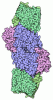
- Assembly
| Deposited unit | 
|
|---|---|
| 1 |
|
-Components
-Nitrogenase MoFe protein ... , 2 types, 4 molecules BDAC
| #1: Protein | Mass: 57094.352 Da / Num. of mol.: 2 / Source method: isolated from a natural source Source: (natural) Gluconacetobacter diazotrophicus PA1 5 (bacteria)References: UniProt: A9H5W8, nitrogenase #2: Protein | Mass: 56095.453 Da / Num. of mol.: 2 / Source method: isolated from a natural source Source: (natural) Gluconacetobacter diazotrophicus PA1 5 (bacteria)References: UniProt: A9H5W5, nitrogenase |
|---|
-Non-polymers , 4 types, 8 molecules 
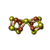
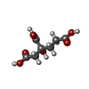
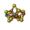
| #3: Chemical | 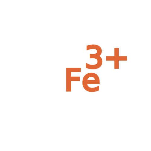 Mass: 55.845 Da / Num. of mol.: 2 / Source method: obtained synthetically / Formula: Fe / Feature type: SUBJECT OF INVESTIGATION Mass: 55.845 Da / Num. of mol.: 2 / Source method: obtained synthetically / Formula: Fe / Feature type: SUBJECT OF INVESTIGATION#4: Chemical | 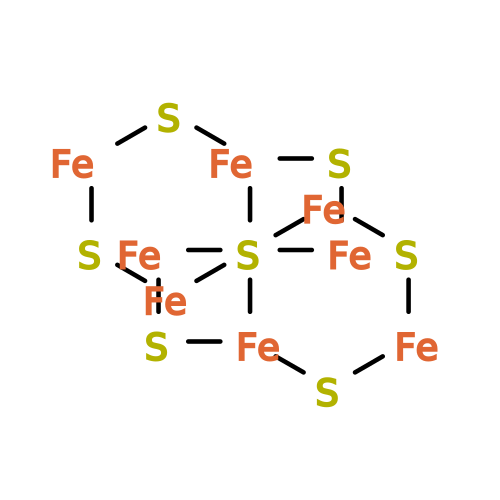 Mass: 671.215 Da / Num. of mol.: 2 / Source method: obtained synthetically / Formula: Fe8S7 / Feature type: SUBJECT OF INVESTIGATION Mass: 671.215 Da / Num. of mol.: 2 / Source method: obtained synthetically / Formula: Fe8S7 / Feature type: SUBJECT OF INVESTIGATION#5: Chemical | 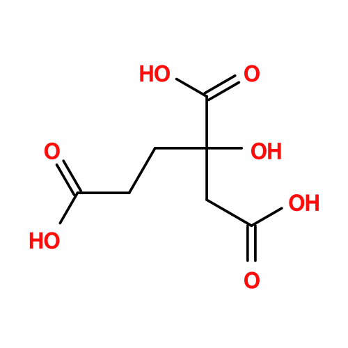 Mass: 206.150 Da / Num. of mol.: 2 / Source method: obtained synthetically / Formula: C7H10O7 / Feature type: SUBJECT OF INVESTIGATION Mass: 206.150 Da / Num. of mol.: 2 / Source method: obtained synthetically / Formula: C7H10O7 / Feature type: SUBJECT OF INVESTIGATION#6: Chemical | 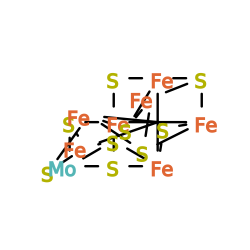 Mass: 787.451 Da / Num. of mol.: 2 / Source method: obtained synthetically / Formula: CFe7MoS9 / Feature type: SUBJECT OF INVESTIGATION Mass: 787.451 Da / Num. of mol.: 2 / Source method: obtained synthetically / Formula: CFe7MoS9 / Feature type: SUBJECT OF INVESTIGATION |
|---|
-Details
| Has ligand of interest | Y |
|---|---|
| Has protein modification | Y |
-Experimental details
-Experiment
| Experiment | Method: ELECTRON MICROSCOPY |
|---|---|
| EM experiment | Aggregation state: PARTICLE / 3D reconstruction method: single particle reconstruction |
- Sample preparation
| Component | Name: Heterotetrameric MoFeP from Gluconacetobacter diazotrophicus Type: COMPLEX / Entity ID: #1-#2 / Source: NATURAL | ||||||||||||||||||||
|---|---|---|---|---|---|---|---|---|---|---|---|---|---|---|---|---|---|---|---|---|---|
| Molecular weight | Value: 0.23 MDa / Experimental value: NO | ||||||||||||||||||||
| Source (natural) | Organism: Gluconacetobacter diazotrophicus PA1 5 (bacteria) | ||||||||||||||||||||
| Buffer solution | pH: 8 Details: Solutions were prepared, filtered, and degassed to Ar immediately prior to the experiment. | ||||||||||||||||||||
| Buffer component |
| ||||||||||||||||||||
| Specimen | Conc.: 6.9 mg/ml / Embedding applied: NO / Shadowing applied: NO / Staining applied: NO / Vitrification applied: YES Details: Homogeneous Gd-MoFeP purified from a gel filtration column(SEC). | ||||||||||||||||||||
| Specimen support | Grid material: COPPER / Grid mesh size: 300 divisions/in. / Grid type: Quantifoil Active R1.2/0.8 | ||||||||||||||||||||
| Vitrification | Instrument: SPT LABTECH CHAMELEON / Cryogen name: ETHANE / Humidity: 75 % / Chamber temperature: 298 K Details: Samples were covered by Al's oil and frozen with the SPT Labtech chameleon. |
- Electron microscopy imaging
| Experimental equipment |  Model: Titan Krios / Image courtesy: FEI Company |
|---|---|
| Microscopy | Model: TFS KRIOS |
| Electron gun | Electron source:  FIELD EMISSION GUN / Accelerating voltage: 300 kV / Illumination mode: FLOOD BEAM FIELD EMISSION GUN / Accelerating voltage: 300 kV / Illumination mode: FLOOD BEAM |
| Electron lens | Mode: BRIGHT FIELD / Nominal magnification: 130000 X / Nominal defocus max: 2500 nm / Nominal defocus min: 1000 nm / Cs: 2.7 mm / Alignment procedure: COMA FREE |
| Specimen holder | Cryogen: NITROGEN / Specimen holder model: FEI TITAN KRIOS AUTOGRID HOLDER |
| Image recording | Average exposure time: 5 sec. / Electron dose: 60 e/Å2 / Film or detector model: FEI FALCON IV (4k x 4k) / Num. of grids imaged: 1 / Num. of real images: 2371 |
| EM imaging optics | Energyfilter name: TFS Selectris X / Energyfilter slit width: 10 eV |
| Image scans | Width: 4096 / Height: 4096 |
- Processing
| EM software |
| ||||||||||||||||||||||||
|---|---|---|---|---|---|---|---|---|---|---|---|---|---|---|---|---|---|---|---|---|---|---|---|---|---|
| CTF correction | Type: PHASE FLIPPING AND AMPLITUDE CORRECTION | ||||||||||||||||||||||||
| Particle selection | Num. of particles selected: 674977 | ||||||||||||||||||||||||
| Symmetry | Point symmetry: C2 (2 fold cyclic) | ||||||||||||||||||||||||
| 3D reconstruction | Resolution: 2.15 Å / Resolution method: FSC 0.143 CUT-OFF / Num. of particles: 211731 / Algorithm: FOURIER SPACE / Symmetry type: POINT | ||||||||||||||||||||||||
| Atomic model building | Protocol: RIGID BODY FIT / Space: REAL | ||||||||||||||||||||||||
| Refinement | Highest resolution: 2.15 Å Stereochemistry target values: REAL-SPACE (WEIGHTED MAP SUM AT ATOM CENTERS) | ||||||||||||||||||||||||
| Refine LS restraints |
|
