 Movie
Movie Controller

[English] 日本語
 Yorodumi
- EMDB-75567: CryoEM structure of Gluconacetobacter diazotrophicus MoFeP (C2 sy... -
+ Open data

- Basic information
| Entry |  | ||||||||||||
|---|---|---|---|---|---|---|---|---|---|---|---|---|---|
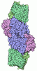
| Title | CryoEM structure of Gluconacetobacter diazotrophicus MoFeP (C2 symmetry) | ||||||||||||
 Map data Map data | Gluconacetobacter diazotrophicus MoFeP (C2 symmetry) sharp map | ||||||||||||
 Sample Sample |
| ||||||||||||
 Keywords Keywords | Nitrogenase / MoFeP / Gluconacetobacter diazotrophicus / molybdenum-iron protein / Oxidoreductase | ||||||||||||
| Function / homology |  Function and homology information Function and homology informationmolybdenum-iron nitrogenase complex / nitrogenase / nitrogenase activity / iron-sulfur cluster binding / ATP binding / metal ion binding Similarity search - Function | ||||||||||||
| Biological species |  Gluconacetobacter diazotrophicus PA1 5 (bacteria) Gluconacetobacter diazotrophicus PA1 5 (bacteria) | ||||||||||||
| Method | single particle reconstruction / cryo EM / Resolution: 2.15 Å | ||||||||||||
 Authors Authors | Li Y / Narehood SM / Cook BD / McGuire KL / Tezcan FA / Herzik Jr MA | ||||||||||||
| Funding support |  United States, 3 items United States, 3 items
| ||||||||||||
 Citation Citation | Journal: Biochemistry / Year: 2026 Title: Structural and Functional Characterization of Heterologous Nitrogenase Complexes. Authors: Yizhou Li / Sarah M Narehood / Brian D Cook / Kelly L McGuire / Mark A Herzik / F Akif Tezcan / Abstract: Nitrogenase is the only known enzyme that catalyzes the reduction of dinitrogen to ammonia. The most prevalent isozyme, molybdenum nitrogenase, comprises the catalytic molybdenum-iron protein (MoFeP) ...Nitrogenase is the only known enzyme that catalyzes the reduction of dinitrogen to ammonia. The most prevalent isozyme, molybdenum nitrogenase, comprises the catalytic molybdenum-iron protein (MoFeP) and the ATP-dependent reductase iron protein (FeP). Although Mo-nitrogenases are widespread across bacteria and archaea and appear to share conserved mechanistic and structural features, FeP and MoFeP show considerable sequence variability across diazotrophs. This raises questions about the conservation of chemomechanical mechanisms coupling FeP-dependent ATP hydrolysis and electron transfer to MoFeP, and about the functional compatibility of nitrogenase components from divergent species. Previous studies showed that some heterologous FeP-MoFeP pairs can functionally complement each other, whereas other pairs lack catalytic activity, but the absence of structural information on such heterologous pairs has limited mechanistic understanding. To this end, we investigated the functional and structural compatibility of FeP and MoFeP from () and (), two phylogenetically and ecologically distinct species. Building on our prior work with -nitrogenase and recently developed cryogenic electron microscopy (cryoEM) protocols, we determined the ADP·BeF-trapped structure of the homologous FeP-MoFeP complex and showed that it adopted the same geometry as its counterpart. Activity measurements showed that heterologous combinations retained 60-80% of homologous catalytic activities despite 30-50% sequence divergence in FeP and MoFeP. High-resolution cryoEM structures of FeP-MoFeP and FeP-MoFeP corroborated these activities and revealed that functional complementation tolerates substantial sequence variation when the core structural elements supporting ATP binding/hydrolysis, protein-protein interaction, electron transfer, and substrate reduction are conserved. | ||||||||||||
| History |
|
- Structure visualization
| Supplemental images |
|---|

- Downloads & links
-EMDB archive
| Map data | emd_75567.map.gz | 323.9 MB | EMDB map data format | |
|---|---|---|---|---|
| Header (meta data) | emd-75567-v30.xmlemd-75567.xml | 29.6 KB 29.6 KB | Display Display | EMDB header |
| FSC (resolution estimation) | emd_75567_fsc.xml | 14.7 KB | Display | FSC data file |
| Images |  emd_75567.png emd_75567.png | 31.5 KB | ||
| Masks | emd_75567_msk_1.map | 343 MB | Mask map | |
| Filedesc metadata | emd-75567.cif.gz | 7.8 KB | ||
| Others | emd_75567_additional_1.map.gzemd_75567_half_map_1.map.gzemd_75567_half_map_2.map.gz | 173.4 MB 317.9 MB 317.9 MB | ||
| Archive directory |  https://data.pdbj.org/pub/emdb/structures/EMD-75567ftp://data.pdbj.org/pub/emdb/structures/EMD-75567 https://data.pdbj.org/pub/emdb/structures/EMD-75567ftp://data.pdbj.org/pub/emdb/structures/EMD-75567 | HTTPS FTP |
-Related structure data
| Related structure data |  10zlMC  10zkC  10zmC  10znC M: atomic model generated by this map C: citing same article ( |
|---|---|
| Similar structure data |


-Links
| EMDB pages | EMDB (EBI/PDBe) / EMDataResource |
|---|---|
| Related items in Molecule of the Month |
-Map
| File | Download / File: emd_75567.map.gz / Format: CCP4 / Size: 343 MB / Type: IMAGE STORED AS FLOATING POINT NUMBER (4 BYTES) | ||||||||||||||||||||||||||||||||||||
|---|---|---|---|---|---|---|---|---|---|---|---|---|---|---|---|---|---|---|---|---|---|---|---|---|---|---|---|---|---|---|---|---|---|---|---|---|---|
| Annotation | Gluconacetobacter diazotrophicus MoFeP (C2 symmetry) sharp map | ||||||||||||||||||||||||||||||||||||
| Projections & slices | Image control
Images are generated by Spider. | ||||||||||||||||||||||||||||||||||||
| Voxel size | X=Y=Z: 0.935 Å | ||||||||||||||||||||||||||||||||||||
| Density |
| ||||||||||||||||||||||||||||||||||||
| Symmetry | Space group: 1 | ||||||||||||||||||||||||||||||||||||
| Details | EMDB XML:
|
 Z (Sec.)
 Y (Row.)
 X (Col.)


-Supplemental data
-Mask #1
| File | emd_75567_msk_1.map | ||||||||||||
|---|---|---|---|---|---|---|---|---|---|---|---|---|---|
| Projections & Slices |
| ||||||||||||
| Density Histograms |


-Additional map: Gluconacetobacter diazotrophicus MoFeP (C2 symmetry) map
| File | emd_75567_additional_1.map | ||||||||||||
|---|---|---|---|---|---|---|---|---|---|---|---|---|---|
| Annotation | Gluconacetobacter diazotrophicus MoFeP (C2 symmetry) map | ||||||||||||
| Projections & Slices |
| ||||||||||||
| Density Histograms |


-Half map: Gluconacetobacter diazotrophicus MoFeP (C2 symmetry) half map
| File | emd_75567_half_map_1.map | ||||||||||||
|---|---|---|---|---|---|---|---|---|---|---|---|---|---|
| Annotation | Gluconacetobacter diazotrophicus MoFeP (C2 symmetry) half map | ||||||||||||
| Projections & Slices |
| ||||||||||||
| Density Histograms |


-Half map: Gluconacetobacter diazotrophicus MoFeP (C2 symmetry) half map
| File | emd_75567_half_map_2.map | ||||||||||||
|---|---|---|---|---|---|---|---|---|---|---|---|---|---|
| Annotation | Gluconacetobacter diazotrophicus MoFeP (C2 symmetry) half map | ||||||||||||
| Projections & Slices |
| ||||||||||||
| Density Histograms |


- Sample components
-Entire : Heterotetrameric MoFeP from Gluconacetobacter diazotrophicus
| Entire | Name: Heterotetrameric MoFeP from Gluconacetobacter diazotrophicus |
|---|---|
| Components |
|
-Supramolecule #1: Heterotetrameric MoFeP from Gluconacetobacter diazotrophicus
| Supramolecule | Name: Heterotetrameric MoFeP from Gluconacetobacter diazotrophicus type: complex / ID: 1 / Parent: 0 / Macromolecule list: #1-#2 |
|---|---|
| Source (natural) | Organism: Gluconacetobacter diazotrophicus PA1 5 (bacteria) |
| Molecular weight | Theoretical: 230 KDa |
-Macromolecule #1: Nitrogenase MoFe protein beta subunit NifK
| Macromolecule | Name: Nitrogenase MoFe protein beta subunit NifK / type: protein_or_peptide / ID: 1 / Number of copies: 2 / Enantiomer: LEVO / EC number: nitrogenase |
|---|---|
| Source (natural) | Organism: Gluconacetobacter diazotrophicus PA1 5 (bacteria) |
| Molecular weight | Theoretical: 57.094352 KDa |
| Sequence | String: MPQNVDKILD HAPLFREPEY QEMLAGKAKL ENMPPADKVV EIADWTKSWE YREKNFARES LSVNPAKACQ PLGAVFVASG FERTMSFVH GSQGCVAYYR SHLSRHFKEP SSAVSSSMTE DAAVFGGLNN MVDGLANTYK LYDPKMIAVS TTCMAEVIGD D LHAFIQTA ...String: MPQNVDKILD HAPLFREPEY QEMLAGKAKL ENMPPADKVV EIADWTKSWE YREKNFARES LSVNPAKACQ PLGAVFVASG FERTMSFVH GSQGCVAYYR SHLSRHFKEP SSAVSSSMTE DAAVFGGLNN MVDGLANTYK LYDPKMIAVS TTCMAEVIGD D LHAFIQTA KGKGSVPEEF DVPFAHTPAF VGSHVTGYDN MLKGILEHFW KGRTPVPNRS VNIIPGFDGF AVGNNRELKR IL GMMGVQY TILSDVSDQF DTPSDGEYRM YDGGTKIEAA RDAVNADYTI SLQEYCTPKT LEYCQSFGQK TASFHYPLGI GAT DDLLQK LSEISGKPVP QELEMERGRL VDALADSQAY LHGKTYAIYG DPDFVYGMAR FILETGGEPK HCLATNGSKA WEAQ MQELF DSSPFGVGCK AWGGKDLWHM RSLLATEKVD LLIGNSYGKY LERDTDTPLI RLMFPIFDRH HHHRFPVWGY QGALR VLVT LLDKIFDKLD DDTIQAGVTD YSFDLTR UniProtKB: Nitrogenase molybdenum-iron protein beta chain |
-Macromolecule #2: Nitrogenase MoFe protein alpha subunit NifD
| Macromolecule | Name: Nitrogenase MoFe protein alpha subunit NifD / type: protein_or_peptide / ID: 2 / Number of copies: 2 / Enantiomer: LEVO / EC number: nitrogenase |
|---|---|
| Source (natural) | Organism: Gluconacetobacter diazotrophicus PA1 5 (bacteria) |
| Molecular weight | Theoretical: 56.095453 KDa |
| Sequence | String: MSLDEDKTND SAFHARLIAE VLEAYPDKAR KRRQKHLNVA GQAEAEAQDA GEEGVMLSEC DVKSNVKSVP GVMTIRGCAY AGSKGVVWG PVKDMVHISH GPVGCGQYSW SQRRNYYIGN TGVDSFVTMQ FTSDFQEKDI VFGGDKKLEK IIDEIDELFP L AKGISVQS ...String: MSLDEDKTND SAFHARLIAE VLEAYPDKAR KRRQKHLNVA GQAEAEAQDA GEEGVMLSEC DVKSNVKSVP GVMTIRGCAY AGSKGVVWG PVKDMVHISH GPVGCGQYSW SQRRNYYIGN TGVDSFVTMQ FTSDFQEKDI VFGGDKKLEK IIDEIDELFP L AKGISVQS ECPIGLIGDD IEAVSRKKKK EIGKTIVPVR CEGFRGVSQS LGHHIANDAI RDWVFDGEDK HAAFETTPYD VN VIGDYNI GGDAWSSRIL LEEMGLRVVG NWSGDATLAE IERAPKAKLN LIHCYRSMNY ICRHMEEKYN IPWTEYNFFG PSQ IAASLR KIAALFDEKI QEGAERVIAK YQPLVDAVIE KFRPRLAGKK VMLYVGGLRP RHVVNAYNDL GMEIVGTGYE FGHN DDYQR TGHYVREGTL IYDDVTGYEL EKFIEGIRPD LVGSGIKEKY PVQKMGIPFR QMHSWDYSGP YHGYDGFAIF ARDMD LAIN NPVWSMFKAP WKNAA UniProtKB: Nitrogenase protein alpha chain |
-Macromolecule #3: FE (III) ION
| Macromolecule | Name: FE (III) ION / type: ligand / ID: 3 / Number of copies: 2 / Formula: FE |
|---|---|
| Molecular weight | Theoretical: 55.845 Da |
-Macromolecule #4: FE(8)-S(7) CLUSTER
| Macromolecule | Name: FE(8)-S(7) CLUSTER / type: ligand / ID: 4 / Number of copies: 2 / Formula: CLF |
|---|---|
| Molecular weight | Theoretical: 671.215 Da |
| Chemical component information | 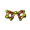 ChemComp-CLF: |
-Macromolecule #5: 3-HYDROXY-3-CARBOXY-ADIPIC ACID
| Macromolecule | Name: 3-HYDROXY-3-CARBOXY-ADIPIC ACID / type: ligand / ID: 5 / Number of copies: 2 / Formula: HCA |
|---|---|
| Molecular weight | Theoretical: 206.15 Da |
| Chemical component information | 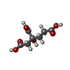 ChemComp-HCA: |
-Macromolecule #6: iron-sulfur-molybdenum cluster with interstitial carbon
| Macromolecule | Name: iron-sulfur-molybdenum cluster with interstitial carbon type: ligand / ID: 6 / Number of copies: 2 / Formula: ICS |
|---|---|
| Molecular weight | Theoretical: 787.451 Da |
| Chemical component information | 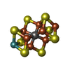 ChemComp-ICE: |
-Experimental details
-Structure determination
| Method | cryo EM |
|---|---|
 Processing Processing | single particle reconstruction |
| Aggregation state | particle |
-Sample preparation
| Concentration | 6.9 mg/mL | ||||||||||||
|---|---|---|---|---|---|---|---|---|---|---|---|---|---|
| Buffer | pH: 8 Component:
Details: Solutions were prepared, filtered, and degassed to Ar immediately prior to the experiment. | ||||||||||||
| Grid | Model: Quantifoil Active R1.2/0.8 / Material: COPPER / Mesh: 300 / Support film - Material: CARBON / Support film - topology: HOLEY / Pretreatment - Type: GLOW DISCHARGE / Pretreatment - Time: 45 sec. | ||||||||||||
| Vitrification | Cryogen name: ETHANE / Chamber humidity: 75 % / Chamber temperature: 298 K / Instrument: SPT LABTECH CHAMELEON Details: Samples were covered by Al's oil and frozen with the SPT Labtech chameleon.. | ||||||||||||
| Details | Homogeneous Gd-MoFeP purified from a gel filtration column(SEC). |
- Electron microscopy
| Microscope | TFS KRIOS |
|---|---|
| Specialist optics | Energy filter - Name: TFS Selectris X / Energy filter - Slit width: 10 eV |
| Image recording | Film or detector model: FEI FALCON IV (4k x 4k) / Digitization - Dimensions - Width: 4096 pixel / Digitization - Dimensions - Height: 4096 pixel / Number grids imaged: 1 / Number real images: 2371 / Average exposure time: 5.0 sec. / Average electron dose: 60.0 e/Å2 |
| Electron beam | Acceleration voltage: 300 kV / Electron source:  FIELD EMISSION GUN FIELD EMISSION GUN |
| Electron optics | Illumination mode: FLOOD BEAM / Imaging mode: BRIGHT FIELD / Cs: 2.7 mm / Nominal defocus max: 2.5 µm / Nominal defocus min: 1.0 µm / Nominal magnification: 130000 |
| Sample stage | Specimen holder model: FEI TITAN KRIOS AUTOGRID HOLDER / Cooling holder cryogen: NITROGEN |
| Experimental equipment |  Model: Titan Krios / Image courtesy: FEI Company |
+Image processing


-Atomic model buiding 1
| Refinement | Space: REAL / Protocol: RIGID BODY FIT |
|---|---|
| Output model | PDB-10zl: |
