Flagellar hook protein FlgE superfamily / Flagellar hook protein FlgE / Flagellar basal body protein FlaE D2 domain / Flagellar hook-basal body protein, FlgE/F/G / Flagellar hook-basal body protein, FlgE/F/G-like / : / Flagellar hook protein FlgE/F/G D1 domain / Flagellar basal body rod protein, conserved site / Flagella basal body rod proteins signature. / Flagellar basal body rod protein, N-terminal ...Flagellar hook protein FlgE superfamily / Flagellar hook protein FlgE / Flagellar basal body protein FlaE D2 domain / Flagellar hook-basal body protein, FlgE/F/G / Flagellar hook-basal body protein, FlgE/F/G-like / : / Flagellar hook protein FlgE/F/G D1 domain / Flagellar basal body rod protein, conserved site / Flagella basal body rod proteins signature. / Flagellar basal body rod protein, N-terminal / Flagellar basal-body/hook protein, C-terminal domain / Flagella basal body rod protein / Flagellar basal body rod FlgEFG protein C-terminal Similarity search - Domain/homology
Japan Agency for Medical Research and Development (AMED)
JP19am0101117
Japan
Japan Agency for Medical Research and Development (AMED)
JP17pc0101020
Japan
Citation
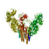
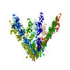
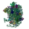
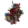
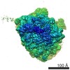
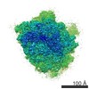
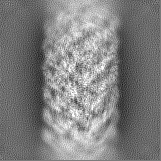
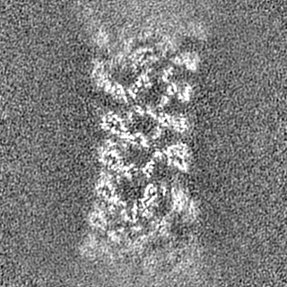
Journal: Nat Commun / Year: 2019 Title: Structure of the native supercoiled flagellar hook as a universal joint. Authors: Takayuki Kato / Fumiaki Makino / Tomoko Miyata / Péter Horváth / Keiichi Namba / Abstract: The Bacterial flagellar hook is a short supercoiled tubular structure made from a helical assembly of the hook protein FlgE. The hook acts as a universal joint that connects the flagellar basal body ...The Bacterial flagellar hook is a short supercoiled tubular structure made from a helical assembly of the hook protein FlgE. The hook acts as a universal joint that connects the flagellar basal body and filament, and smoothly transmits torque generated by the rotary motor to the helical filament propeller. In peritrichously flagellated bacteria, the hook allows the filaments to form a bundle behind the cell for swimming, and for the bundle to fall apart for tumbling. Here we report a native supercoiled hook structure at 3.6 Å resolution by cryoEM single particle image analysis of the polyhook. The atomic model built into the three-dimensional (3D) density map reveals the changes in subunit conformation and intersubunit interactions that occur upon compression and extension of the 11 protofilaments during their smoke ring-like rotation. These observations reveal how the hook functions as a dynamic molecular universal joint with high bending flexibility and twisting rigidity.
History
Deposition
Jun 17, 2019
-
Header (metadata) release
Feb 12, 2020
-
Map release
Feb 12, 2020
-
Update
Mar 27, 2024
-
Current status
Mar 27, 2024
Processing site: PDBj / Status: Released
-
Structure visualization
Movie
Surface view with section colored by density value
In the structure databanks used in Yorodumi, some data are registered as the other names, "COVID-19 virus" and "2019-nCoV". Here are the details of the virus and the list of structure data.
Jan 31, 2019. EMDB accession codes are about to change! (news from PDBe EMDB page)
EMDB accession codes are about to change! (news from PDBe EMDB page)
The allocation of 4 digits for EMDB accession codes will soon come to an end. Whilst these codes will remain in use, new EMDB accession codes will include an additional digit and will expand incrementally as the available range of codes is exhausted. The current 4-digit format prefixed with “EMD-” (i.e. EMD-XXXX) will advance to a 5-digit format (i.e. EMD-XXXXX), and so on. It is currently estimated that the 4-digit codes will be depleted around Spring 2019, at which point the 5-digit format will come into force.
The EM Navigator/Yorodumi systems omit the EMD- prefix.
Related info.:Q: What is EMD? / ID/Accession-code notation in Yorodumi/EM Navigator
Yorodumi is a browser for structure data from EMDB, PDB, SASBDB, etc.
This page is also the successor to EM Navigator detail page, and also detail information page/front-end page for Omokage search.
The word "yorodu" (or yorozu) is an old Japanese word meaning "ten thousand". "mi" (miru) is to see.
Related info.:EMDB / PDB / SASBDB / Comparison of 3 databanks / Yorodumi Search / Aug 31, 2016. New EM Navigator & Yorodumi / Yorodumi Papers / Jmol/JSmol / Function and homology information / Changes in new EM Navigator and Yorodumi
 Movie
Movie Controller


 Open data
 Basic information
 Map data
 Sample
 Keywords
 Function and homology information
 Salmonella enterica subsp. enterica serovar Typhimurium (bacteria) /
  Authors
 Japan, 4 items
  Citation
 Structure visualization
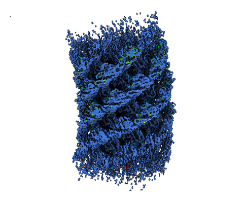
 Downloads & links
 emd_9952.png
 http://ftp.pdbj.org/pub/emdb/structures/EMD-9952
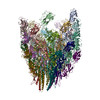


 Z (Sec.)
 Y (Row.)
 X (Col.)


 Sample components
 Processing
 Electron microscopy
 FIELD EMISSION GUN