[English] 日本語
 Yorodumi
- EMDB-9743: Cryo-EM structure of the S. typhimurium oxaloacetate decarboxylas... -
+ Open data
- Basic information
| Entry | Database: EMDB / ID: EMD-9743 | |||||||||
|---|---|---|---|---|---|---|---|---|---|---|
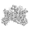
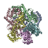
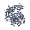
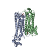
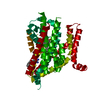
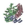
| Title | Cryo-EM structure of the S. typhimurium oxaloacetate decarboxylase beta-gamma sub-complex | |||||||||
 Map data Map data | ||||||||||
 Sample Sample |
| |||||||||
 Keywords Keywords | membrane protein / sodium pump / decarboxylase sodium pump / biotin-dependent decarboxylase | |||||||||
| Function / homology |  Function and homology information Function and homology informationoxaloacetate decarboxylase (Na+ extruding) / decarboxylation-driven active transmembrane transporter activity / sodium ion transmembrane transporter activity / oxaloacetate decarboxylase activity / sodium ion export across plasma membrane / sodium ion transport / lyase activity / plasma membrane Similarity search - Function | |||||||||
| Biological species |  Salmonella enterica subsp. enterica serovar Typhimurium (bacteria) Salmonella enterica subsp. enterica serovar Typhimurium (bacteria) | |||||||||
| Method | single particle reconstruction / cryo EM / Resolution: 3.9 Å | |||||||||
 Authors Authors | Xu X / Shi H | |||||||||
| Funding support |  China, 1 items China, 1 items
| |||||||||
 Citation Citation |  Journal: Elife / Year: 2020 Journal: Elife / Year: 2020Title: Structural insights into sodium transport by the oxaloacetate decarboxylase sodium pump. Authors: Xin Xu / Huigang Shi / Xiaowen Gong / Pu Chen / Ying Gao / Xinzheng Zhang / Song Xiang /  Abstract: The oxaloacetate decarboxylase sodium pump (OAD) is a unique primary-active transporter that utilizes the free energy derived from oxaloacetate decarboxylation for sodium transport across the cell ...The oxaloacetate decarboxylase sodium pump (OAD) is a unique primary-active transporter that utilizes the free energy derived from oxaloacetate decarboxylation for sodium transport across the cell membrane. It is composed of 3 subunits: the α subunit catalyzes carboxyl-transfer from oxaloacetate to biotin, the membrane integrated β subunit catalyzes the subsequent carboxyl-biotin decarboxylation and the coupled sodium transport, the γ subunit interacts with the α and β subunits and stabilizes the OAD complex. We present here structure of the OAD βγ sub-complex. The structure revealed that the β and γ subunits form a βγ hetero-hexamer with extensive interactions between the subunits and shed light on the OAD holo-enzyme assembly. Structure-guided functional studies provided insights into the sodium binding sites in the β subunit and the coupling between carboxyl-biotin decarboxylation and sodium transport by the OAD β subunit. | |||||||||
| History |
|
- Structure visualization
| Movie |
 Movie viewer Movie viewer |
|---|---|
| Structure viewer | EM map:  SurfView SurfView Molmil Molmil Jmol/JSmol Jmol/JSmol |
| Supplemental images |
- Downloads & links
-EMDB archive
| Map data |  emd_9743.map.gz emd_9743.map.gz | 28.6 MB |  EMDB map data format EMDB map data format | |
|---|---|---|---|---|
| Header (meta data) |  emd-9743-v30.xml emd-9743-v30.xml emd-9743.xml emd-9743.xml | 13.5 KB 13.5 KB | Display Display |  EMDB header EMDB header |
| Images |  emd_9743.png emd_9743.png | 63.7 KB | ||
| Filedesc metadata |  emd-9743.cif.gz emd-9743.cif.gz | 6 KB | ||
| Archive directory |  http://ftp.pdbj.org/pub/emdb/structures/EMD-9743 http://ftp.pdbj.org/pub/emdb/structures/EMD-9743 ftp://ftp.pdbj.org/pub/emdb/structures/EMD-9743 ftp://ftp.pdbj.org/pub/emdb/structures/EMD-9743 | HTTPS FTP |
-Validation report
| Summary document |  emd_9743_validation.pdf.gz emd_9743_validation.pdf.gz | 610.9 KB | Display |  EMDB validaton report EMDB validaton report |
|---|---|---|---|---|
| Full document |  emd_9743_full_validation.pdf.gz emd_9743_full_validation.pdf.gz | 610.4 KB | Display | |
| Data in XML |  emd_9743_validation.xml.gz emd_9743_validation.xml.gz | 5.7 KB | Display | |
| Data in CIF |  emd_9743_validation.cif.gz emd_9743_validation.cif.gz | 6.5 KB | Display | |
| Arichive directory |  https://ftp.pdbj.org/pub/emdb/validation_reports/EMD-9743 https://ftp.pdbj.org/pub/emdb/validation_reports/EMD-9743 ftp://ftp.pdbj.org/pub/emdb/validation_reports/EMD-9743 ftp://ftp.pdbj.org/pub/emdb/validation_reports/EMD-9743 | HTTPS FTP |
-Related structure data
| Related structure data |  6iwwMC  6ivaC M: atomic model generated by this map C: citing same article ( |
|---|---|
| Similar structure data |
- Links
Links
| EMDB pages |  EMDB (EBI/PDBe) / EMDB (EBI/PDBe) /  EMDataResource EMDataResource |
|---|
- Map
Map
| File |  Download / File: emd_9743.map.gz / Format: CCP4 / Size: 30.5 MB / Type: IMAGE STORED AS FLOATING POINT NUMBER (4 BYTES) Download / File: emd_9743.map.gz / Format: CCP4 / Size: 30.5 MB / Type: IMAGE STORED AS FLOATING POINT NUMBER (4 BYTES) | ||||||||||||||||||||||||||||||||||||||||||||||||||||||||||||||||||||
|---|---|---|---|---|---|---|---|---|---|---|---|---|---|---|---|---|---|---|---|---|---|---|---|---|---|---|---|---|---|---|---|---|---|---|---|---|---|---|---|---|---|---|---|---|---|---|---|---|---|---|---|---|---|---|---|---|---|---|---|---|---|---|---|---|---|---|---|---|---|
| Projections & slices | Image control
Images are generated by Spider. | ||||||||||||||||||||||||||||||||||||||||||||||||||||||||||||||||||||
| Voxel size | X=Y=Z: 1.3203 Å | ||||||||||||||||||||||||||||||||||||||||||||||||||||||||||||||||||||
| Density |
| ||||||||||||||||||||||||||||||||||||||||||||||||||||||||||||||||||||
| Symmetry | Space group: 1 | ||||||||||||||||||||||||||||||||||||||||||||||||||||||||||||||||||||
| Details | EMDB XML:
CCP4 map header:
| ||||||||||||||||||||||||||||||||||||||||||||||||||||||||||||||||||||
-Supplemental data
- Sample components
-Entire : The oxaloacetate decarboxylase beta-gamma sub-complex
| Entire | Name: The oxaloacetate decarboxylase beta-gamma sub-complex |
|---|---|
| Components |
|
-Supramolecule #1: The oxaloacetate decarboxylase beta-gamma sub-complex
| Supramolecule | Name: The oxaloacetate decarboxylase beta-gamma sub-complex / type: complex / ID: 1 / Parent: 0 / Macromolecule list: #2 |
|---|---|
| Source (natural) | Organism:  Salmonella enterica subsp. enterica serovar Typhimurium (bacteria) Salmonella enterica subsp. enterica serovar Typhimurium (bacteria)Strain: enterica serovar Typhimurium |
-Macromolecule #1: Oxaloacetate decarboxylase beta chain
| Macromolecule | Name: Oxaloacetate decarboxylase beta chain / type: protein_or_peptide / ID: 1 / Number of copies: 3 / Enantiomer: LEVO / EC number: oxaloacetate decarboxylase (Na+ extruding) |
|---|---|
| Source (natural) | Organism:  Salmonella enterica subsp. enterica serovar Typhimurium (bacteria) Salmonella enterica subsp. enterica serovar Typhimurium (bacteria)Strain: enterica serovar Typhimurium |
| Molecular weight | Theoretical: 44.928801 KDa |
| Recombinant expression | Organism:  |
| Sequence | String: MESLNALLQG MGLMHLGAGQ AIMLLVSLLL LWLAIAKKFE PLLLLPIGFG GLLSNIPEAG MALTALESLL AHHDAGQLAV IAAKLNCAP DVHAIKEALA LALPSVQGQM ENLAVDMGYT PGVLALFYKV AIGSGVAPLV IFMGVGAMTD FGPLLANPRT L LLGAAAQF ...String: MESLNALLQG MGLMHLGAGQ AIMLLVSLLL LWLAIAKKFE PLLLLPIGFG GLLSNIPEAG MALTALESLL AHHDAGQLAV IAAKLNCAP DVHAIKEALA LALPSVQGQM ENLAVDMGYT PGVLALFYKV AIGSGVAPLV IFMGVGAMTD FGPLLANPRT L LLGAAAQF GIFATVLGAL TLNYFGLISF TLPQAAAIGI IGGADGPTAI YLSGKLAPEL LGAIAVAAYS YMALVPLIQP PI MRALTSE KERKIRMVQL RTVSKREKIL FPVVLLLLVA LLLPDAAPLL GMFCFGNLMR ESGVVERLSD TVQNGLINIV TIF LGLSVG AKLVADKFLQ PQTLGILLLG VIAFGIGTAA GVLMAKLLNL CSKNKINPLI GSAGVSAVPM AARVSNKVGL ESDA QNFLL MHAMGPNVAG VIGSAIAAGV MLKYVLAM UniProtKB: Oxaloacetate decarboxylase beta chain |
-Macromolecule #2: Probable oxaloacetate decarboxylase gamma chain
| Macromolecule | Name: Probable oxaloacetate decarboxylase gamma chain / type: protein_or_peptide / ID: 2 / Number of copies: 3 / Enantiomer: LEVO / EC number: oxaloacetate decarboxylase (Na+ extruding) |
|---|---|
| Source (natural) | Organism:  Salmonella enterica subsp. enterica serovar Typhimurium (bacteria) Salmonella enterica subsp. enterica serovar Typhimurium (bacteria)Strain: enterica serovar Typhimurium |
| Molecular weight | Theoretical: 11.137029 KDa |
| Recombinant expression | Organism:  |
| Sequence | String: MGSSHHHHHH SSGLVPRGSH MTNAALLLGE GFTLMFLGMG FVLAFLFLLI FAIRGMSAAV NRFFPEPAPA PKAAPAAAAP VVDDFTRLK PVIAAAIHHH HRLNA UniProtKB: Probable oxaloacetate decarboxylase gamma chain |
-Macromolecule #3: DODECYL-BETA-D-MALTOSIDE
| Macromolecule | Name: DODECYL-BETA-D-MALTOSIDE / type: ligand / ID: 3 / Number of copies: 3 / Formula: LMT |
|---|---|
| Molecular weight | Theoretical: 510.615 Da |
| Chemical component information |  ChemComp-LMT: |
-Experimental details
-Structure determination
| Method | cryo EM |
|---|---|
 Processing Processing | single particle reconstruction |
| Aggregation state | particle |
- Sample preparation
| Concentration | 8 mg/mL |
|---|---|
| Buffer | pH: 7.5 |
| Grid | Model: Quantifoil R1.2/1.3 / Material: COPPER / Pretreatment - Type: GLOW DISCHARGE / Pretreatment - Time: 60 sec. |
| Vitrification | Cryogen name: ETHANE |
| Details | This sample was monodisperse |
- Electron microscopy
| Microscope | FEI TITAN KRIOS |
|---|---|
| Image recording | Film or detector model: GATAN K2 SUMMIT (4k x 4k) / Detector mode: SUPER-RESOLUTION / Digitization - Frames/image: 1-50 / Number grids imaged: 1 / Number real images: 821 / Average electron dose: 50.0 e/Å2 |
| Electron beam | Acceleration voltage: 300 kV / Electron source:  FIELD EMISSION GUN FIELD EMISSION GUN |
| Electron optics | Calibrated defocus max: 3.5 µm / Calibrated defocus min: 1.5 µm / Illumination mode: FLOOD BEAM / Imaging mode: BRIGHT FIELD |
| Sample stage | Specimen holder model: FEI TITAN KRIOS AUTOGRID HOLDER / Cooling holder cryogen: NITROGEN |
| Experimental equipment |  Model: Titan Krios / Image courtesy: FEI Company |
 Movie
Movie Controller





 Z (Sec.)
 X (Row.)
 Y (Col.)


