[English] 日本語
 Yorodumi
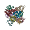
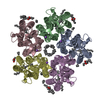
- EMDB-9641: Calcium release-activated calcium channel protein 1, P288L mutant -
+ Open data
- Basic information
| Entry | Database: EMDB / ID: EMD-9641 | |||||||||
|---|---|---|---|---|---|---|---|---|---|---|
| Title | Calcium release-activated calcium channel protein 1, P288L mutant | |||||||||
 Map data Map data | ||||||||||
 Sample Sample |
| |||||||||
| Biological species |  | |||||||||
| Method | single particle reconstruction / cryo EM / Resolution: 5.6 Å | |||||||||
 Authors Authors | Liu X / Wu G / Yang X / Shen Y | |||||||||
 Citation Citation |  Journal: PLoS Biol / Year: 2019 Journal: PLoS Biol / Year: 2019Title: Molecular understanding of calcium permeation through the open Orai channel. Authors: Xiaofen Liu / Guangyan Wu / Yi Yu / Xiaozhe Chen / Renci Ji / Jing Lu / Xin Li / Xing Zhang / Xue Yang / Yuequan Shen /  Abstract: The Orai channel is characterized by voltage independence, low conductance, and high Ca2+ selectivity and plays an important role in Ca2+ influx through the plasma membrane (PM). How the channel is ...The Orai channel is characterized by voltage independence, low conductance, and high Ca2+ selectivity and plays an important role in Ca2+ influx through the plasma membrane (PM). How the channel is activated and promotes Ca2+ permeation is not well understood. Here, we report the crystal structure and cryo-electron microscopy (cryo-EM) reconstruction of a Drosophila melanogaster Orai (dOrai) mutant (P288L) channel that is constitutively active according to electrophysiology. The open state of the Orai channel showed a hexameric assembly in which 6 transmembrane 1 (TM1) helices in the center form the ion-conducting pore, and 6 TM4 helices in the periphery form extended long helices. Orai channel activation requires conformational transduction from TM4 to TM1 and eventually causes the basic section of TM1 to twist outward. The wider pore on the cytosolic side aggregates anions to increase the potential gradient across the membrane and thus facilitate Ca2+ permeation. The open-state structure of the Orai channel offers insights into channel assembly, channel activation, and Ca2+ permeation. | |||||||||
| History |
|
- Structure visualization
| Movie |
 Movie viewer Movie viewer |
|---|---|
| Structure viewer | EM map:  SurfView SurfView Molmil Molmil Jmol/JSmol Jmol/JSmol |
| Supplemental images |
- Downloads & links
-EMDB archive
| Map data |  emd_9641.map.gz emd_9641.map.gz | 1.6 MB |  EMDB map data format EMDB map data format | |
|---|---|---|---|---|
| Header (meta data) |  emd-9641-v30.xml emd-9641-v30.xml emd-9641.xml emd-9641.xml | 10 KB 10 KB | Display Display |  EMDB header EMDB header |
| FSC (resolution estimation) |  emd_9641_fsc.xml emd_9641_fsc.xml | 7.9 KB | Display |  FSC data file FSC data file |
| Images |  emd_9641.png emd_9641.png | 63.3 KB | ||
| Archive directory |  http://ftp.pdbj.org/pub/emdb/structures/EMD-9641 http://ftp.pdbj.org/pub/emdb/structures/EMD-9641 ftp://ftp.pdbj.org/pub/emdb/structures/EMD-9641 ftp://ftp.pdbj.org/pub/emdb/structures/EMD-9641 | HTTPS FTP |
-Related structure data
| Similar structure data |
|---|
- Links
Links
| EMDB pages |  EMDB (EBI/PDBe) / EMDB (EBI/PDBe) /  EMDataResource EMDataResource |
|---|
- Map
Map
| File |  Download / File: emd_9641.map.gz / Format: CCP4 / Size: 40.6 MB / Type: IMAGE STORED AS FLOATING POINT NUMBER (4 BYTES) Download / File: emd_9641.map.gz / Format: CCP4 / Size: 40.6 MB / Type: IMAGE STORED AS FLOATING POINT NUMBER (4 BYTES) | ||||||||||||||||||||||||||||||||||||||||||||||||||||||||||||||||||||
|---|---|---|---|---|---|---|---|---|---|---|---|---|---|---|---|---|---|---|---|---|---|---|---|---|---|---|---|---|---|---|---|---|---|---|---|---|---|---|---|---|---|---|---|---|---|---|---|---|---|---|---|---|---|---|---|---|---|---|---|---|---|---|---|---|---|---|---|---|---|
| Projections & slices | Image control
Images are generated by Spider. | ||||||||||||||||||||||||||||||||||||||||||||||||||||||||||||||||||||
| Voxel size | X=Y=Z: 1.014 Å | ||||||||||||||||||||||||||||||||||||||||||||||||||||||||||||||||||||
| Density |
| ||||||||||||||||||||||||||||||||||||||||||||||||||||||||||||||||||||
| Symmetry | Space group: 1 | ||||||||||||||||||||||||||||||||||||||||||||||||||||||||||||||||||||
| Details | EMDB XML:
CCP4 map header:
| ||||||||||||||||||||||||||||||||||||||||||||||||||||||||||||||||||||
-Supplemental data
- Sample components
-Entire : Calcium release-activated calcium channel protein 1
| Entire | Name: Calcium release-activated calcium channel protein 1 |
|---|---|
| Components |
|
-Supramolecule #1: Calcium release-activated calcium channel protein 1
| Supramolecule | Name: Calcium release-activated calcium channel protein 1 / type: organelle_or_cellular_component / ID: 1 / Parent: 0 / Macromolecule list: #1 |
|---|---|
| Source (natural) | Organism:  |
| Recombinant expression | Organism:  Homo sapiens (human) / Recombinant cell: HEK293S-GnTi- / Recombinant plasmid: pEG-BacMam Homo sapiens (human) / Recombinant cell: HEK293S-GnTi- / Recombinant plasmid: pEG-BacMam |
-Experimental details
-Structure determination
| Method | cryo EM |
|---|---|
 Processing Processing | single particle reconstruction |
| Aggregation state | particle |
- Sample preparation
| Buffer | pH: 8 |
|---|---|
| Grid | Material: COPPER |
| Vitrification | Cryogen name: ETHANE / Chamber humidity: 100 % / Chamber temperature: 293 K / Instrument: FEI VITROBOT MARK IV |
- Electron microscopy
| Microscope | FEI TITAN KRIOS |
|---|---|
| Image recording | Film or detector model: GATAN K2 SUMMIT (4k x 4k) / Detector mode: SUPER-RESOLUTION / Average electron dose: 49.8 e/Å2 |
| Electron beam | Acceleration voltage: 300 kV / Electron source:  FIELD EMISSION GUN FIELD EMISSION GUN |
| Electron optics | Illumination mode: OTHER / Imaging mode: BRIGHT FIELD |
| Sample stage | Specimen holder model: FEI TITAN KRIOS AUTOGRID HOLDER / Cooling holder cryogen: NITROGEN |
| Experimental equipment |  Model: Titan Krios / Image courtesy: FEI Company |
 Movie
Movie Controller


 Z (Sec.)
 Y (Row.)
 X (Col.)


