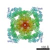
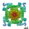
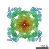
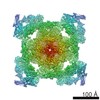
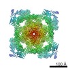
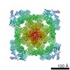
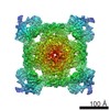
Journal: PLoS Pathog / Year: 2018 Title: Different functional states of fusion protein gB revealed on human cytomegalovirus by cryo electron tomography with Volta phase plate. Authors: Zhu Si / Jiayan Zhang / Sakar Shivakoti / Ivo Atanasov / Chang-Lu Tao / Wong H Hui / Kang Zhou / Xuekui Yu / Weike Li / Ming Luo / Guo-Qiang Bi / Z Hong Zhou / Abstract: Human cytomegalovirus (HCMV) enters host by glycoprotein B (gB)-mediated membrane fusion upon receptor-binding to gH/gL-related complexes, causing devastating diseases such as birth defects. Although ...Human cytomegalovirus (HCMV) enters host by glycoprotein B (gB)-mediated membrane fusion upon receptor-binding to gH/gL-related complexes, causing devastating diseases such as birth defects. Although an X-ray crystal structure of the recombinant gB ectodomain at postfusion conformation is available, the structures of prefusion gB and its complex with gH/gL on the viral envelope remain elusive. Here, we demonstrate the utility of cryo electron tomography (cryoET) with energy filtering and the cutting-edge technologies of Volta phase plate (VPP) and direct electron-counting detection to capture metastable prefusion viral fusion proteins and report the structures of glycoproteins in the native environment of HCMV virions. We established the validity of our approach by obtaining cryoET in situ structures of the vesicular stomatitis virus (VSV) glycoprotein G trimer (171 kD) in prefusion and postfusion conformations, which agree with the known crystal structures of purified G trimers in both conformations. The excellent contrast afforded by these technologies has enabled us to identify gB trimers (303kD) in two distinct conformations in HCMV tomograms and obtain their in situ structures at up to 21 Å resolution through subtomographic averaging. The predominant conformation (79%), which we designate as gB prefusion conformation, fashions a globular endodomain and a Christmas tree-shaped ectodomain, while the minority conformation (21%) has a columnar tree-shaped ectodomain that matches the crystal structure of the "postfusion" gB ectodomain. We also observed prefusion gB in complex with an "L"-shaped density attributed to the gH/gL complex. Integration of these structures of HCMV glycoproteins in multiple functional states and oligomeric forms with existing biochemical data and domain organization of other class III viral fusion proteins suggests that gH/gL receptor-binding triggers conformational changes of gB endodomain, which in turn triggers two essential steps to actuate virus-cell membrane fusion: exposure of gB fusion loops and unfurling of gB ectodomain.
History
Deposition
Nov 12, 2018
-
Header (metadata) release
Nov 28, 2018
-
Map release
Nov 28, 2018
-
Update
Dec 19, 2018
-
Current status
Dec 19, 2018
Processing site: RCSB / Status: Released
-
Structure visualization
Movie
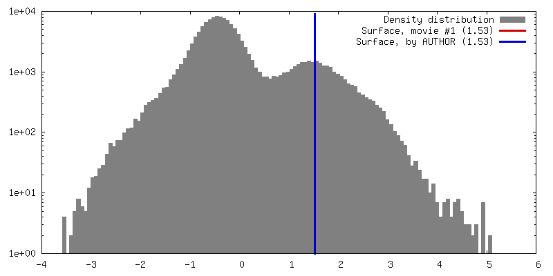
Surface view with section colored by density value
In the structure databanks used in Yorodumi, some data are registered as the other names, "COVID-19 virus" and "2019-nCoV". Here are the details of the virus and the list of structure data.
Jan 31, 2019. EMDB accession codes are about to change! (news from PDBe EMDB page)
EMDB accession codes are about to change! (news from PDBe EMDB page)
The allocation of 4 digits for EMDB accession codes will soon come to an end. Whilst these codes will remain in use, new EMDB accession codes will include an additional digit and will expand incrementally as the available range of codes is exhausted. The current 4-digit format prefixed with “EMD-” (i.e. EMD-XXXX) will advance to a 5-digit format (i.e. EMD-XXXXX), and so on. It is currently estimated that the 4-digit codes will be depleted around Spring 2019, at which point the 5-digit format will come into force.
The EM Navigator/Yorodumi systems omit the EMD- prefix.
Related info.:Q: What is EMD? / ID/Accession-code notation in Yorodumi/EM Navigator
Yorodumi is a browser for structure data from EMDB, PDB, SASBDB, etc.
This page is also the successor to EM Navigator detail page, and also detail information page/front-end page for Omokage search.
The word "yorodu" (or yorozu) is an old Japanese word meaning "ten thousand". "mi" (miru) is to see.
Related info.:EMDB / PDB / SASBDB / Comparison of 3 databanks / Yorodumi Search / Aug 31, 2016. New EM Navigator & Yorodumi / Yorodumi Papers / Jmol/JSmol / Function and homology information / Changes in new EM Navigator and Yorodumi
 Movie
Movie Controller

 Yorodumi
 Open data
 Basic information
 Map data
 Sample
 Human herpesvirus 5 strain AD169
 Authors
 Citation

 Structure visualization
 Movie viewer
 Downloads & links
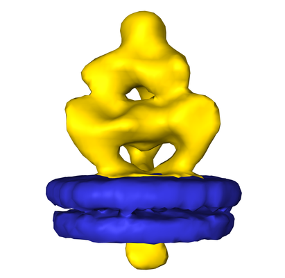 emd_9328.png
 http://ftp.pdbj.org/pub/emdb/structures/EMD-9328

 Z (Sec.)
 Y (Row.)
 X (Col.)


 Sample components
 Processing
 Electron microscopy
 FIELD EMISSION GUN
