National Institutes of Health/National Human Genome Research Institute (NIH/NHGRI)
1R01GM123089
United States
National Institutes of Health/National Human Genome Research Institute (NIH/NHGRI)
1R01GM124348-01
United States
National Institutes of Health/National Human Genome Research Institute (NIH/NHGRI)
R01GM097194
United States
American Heart Association
18POST34030308
United States
German Research Foundation (DFG)
AR1164/1-1
Germany
Citation
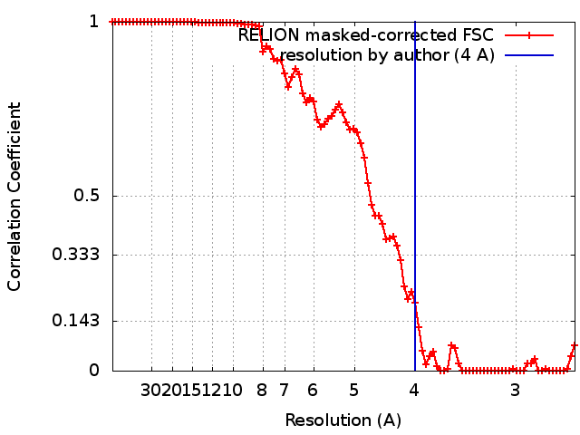
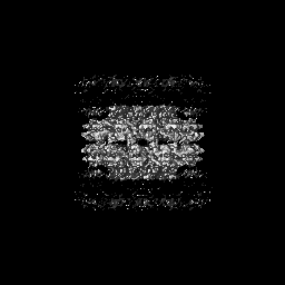
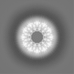
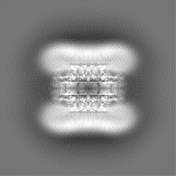
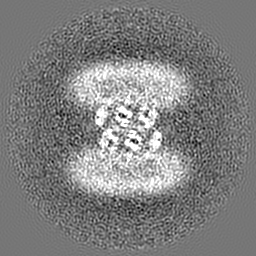
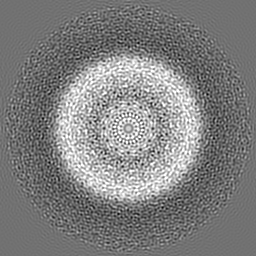
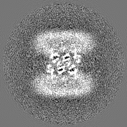
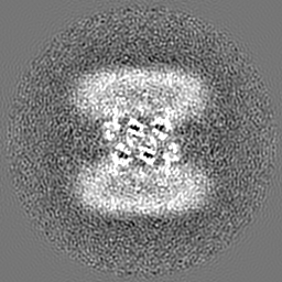
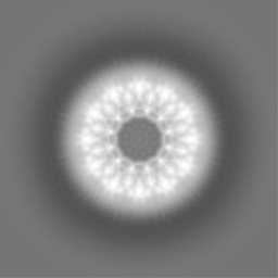
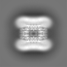
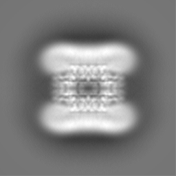
Journal: J Cell Biol / Year: 2018 Title: Cryo-electron microscopy structure of the lipid droplet-formation protein seipin. Authors: Xuewu Sui / Henning Arlt / Kelly P Brock / Zon Weng Lai / Frank DiMaio / Debora S Marks / Maofu Liao / Robert V Farese / Tobias C Walther / Abstract: Metabolic energy is stored in cells primarily as triacylglycerols in lipid droplets (LDs), and LD dysregulation leads to metabolic diseases. The formation of monolayer-bound LDs from the endoplasmic ...Metabolic energy is stored in cells primarily as triacylglycerols in lipid droplets (LDs), and LD dysregulation leads to metabolic diseases. The formation of monolayer-bound LDs from the endoplasmic reticulum (ER) bilayer is poorly understood, but the ER protein seipin is essential to this process. In this study, we report a cryo-electron microscopy structure and functional characterization of seipin. The structure reveals a ring-shaped dodecamer with the luminal domain of each monomer resolved at ∼4.0 Å. Each luminal domain monomer exhibits two distinctive features: a hydrophobic helix (HH) positioned toward the ER bilayer and a β-sandwich domain with structural similarity to lipid-binding proteins. This structure and our functional testing in cells suggest a model in which seipin oligomers initially detect forming LDs in the ER via HHs and subsequently act as membrane anchors to enable lipid transfer and LD growth.
History
Deposition
Sep 28, 2018
-
Header (metadata) release
Oct 17, 2018
-
Map release
Oct 17, 2018
-
Update
Jun 4, 2025
-
Current status
Jun 4, 2025
Processing site: RCSB / Status: Released
-
Structure visualization
Movie
Surface view with section colored by density value
In the structure databanks used in Yorodumi, some data are registered as the other names, "COVID-19 virus" and "2019-nCoV". Here are the details of the virus and the list of structure data.
Jan 31, 2019. EMDB accession codes are about to change! (news from PDBe EMDB page)
EMDB accession codes are about to change! (news from PDBe EMDB page)
The allocation of 4 digits for EMDB accession codes will soon come to an end. Whilst these codes will remain in use, new EMDB accession codes will include an additional digit and will expand incrementally as the available range of codes is exhausted. The current 4-digit format prefixed with “EMD-” (i.e. EMD-XXXX) will advance to a 5-digit format (i.e. EMD-XXXXX), and so on. It is currently estimated that the 4-digit codes will be depleted around Spring 2019, at which point the 5-digit format will come into force.
The EM Navigator/Yorodumi systems omit the EMD- prefix.
Related info.:Q: What is EMD? / ID/Accession-code notation in Yorodumi/EM Navigator
Yorodumi is a browser for structure data from EMDB, PDB, SASBDB, etc.
This page is also the successor to EM Navigator detail page, and also detail information page/front-end page for Omokage search.
The word "yorodu" (or yorozu) is an old Japanese word meaning "ten thousand". "mi" (miru) is to see.
Related info.:EMDB / PDB / SASBDB / Comparison of 3 databanks / Yorodumi Search / Aug 31, 2016. New EM Navigator & Yorodumi / Yorodumi Papers / Jmol/JSmol / Function and homology information / Changes in new EM Navigator and Yorodumi
 Movie
Movie Controller

 Yorodumi
 Open data
 Basic information
 Map data
 Sample
 Keywords
 Function and homology information
 Authors
 United States,
  Germany, 6 items
  Citation
 Structure visualization
 Downloads & links
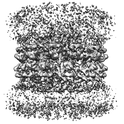 emd_9146.png
 http://ftp.pdbj.org/pub/emdb/structures/EMD-9146

 Z (Sec.)
 Y (Row.)
 X (Col.)


 Sample components
 Processing
 Electron microscopy
 FIELD EMISSION GUN