Journal: J Mol Biol / Year: 2019 Title: Fluorescent Trimeric Hemagglutinins Reveal Multivalent Receptor Binding Properties. Authors: Nikoloz Nemanichvili / Ilhan Tomris / Hannah L Turner / Ryan McBride / Oliver C Grant / Roosmarijn van der Woude / Mohammed H Aldosari / Roland J Pieters / Robert J Woods / James C Paulson / ...Authors: Nikoloz Nemanichvili / Ilhan Tomris / Hannah L Turner / Ryan McBride / Oliver C Grant / Roosmarijn van der Woude / Mohammed H Aldosari / Roland J Pieters / Robert J Woods / James C Paulson / Geert-Jan Boons / Andrew B Ward / Monique H Verheije / Robert P de Vries / Abstract: Influenza A virus carries hundreds of trimeric hemagglutinin (HA) proteins on its viral envelope that interact with various sialylated glycans on a host cell. This interaction represents a ...Influenza A virus carries hundreds of trimeric hemagglutinin (HA) proteins on its viral envelope that interact with various sialylated glycans on a host cell. This interaction represents a multivalent binding event that is present in all the current receptor binding assays, including those employing viruses or precomplexed HA trimers. To study the nature of such multivalent binding events, we fused a superfolder green fluorescent protein (sfGFP) to the C-terminus of trimeric HA to allow for direct visualization of HA-receptor interactions without the need for additional fluorescent antibodies. The multivalent binding of the HA-sfGFP proteins was studied using glycan arrays and tissue staining. The HA-sfGFP with human-type receptor specificity was able to bind to a glycan array as the free trimer. In contrast, the HA-sfGFP with avian-type receptor specificity required multimerization by antibodies before binding to glycans on the glycan array could be observed. Interestingly, multimerization was not required for binding to tissues. The array data may be explained by the possible bivalent binding mode of a single human-specific HA trimer to complex branched N-glycans, which is not possible for the avian-specific HA due to geometrical constrains of the binding sites. The fact that this specificity pattern changes upon interaction with a cell surface probably represents the enhanced amount of glycan orientations and variable densities versus those on the glycan array.
History
Deposition
Aug 27, 2018
-
Header (metadata) release
Sep 19, 2018
-
Map release
Jan 16, 2019
-
Update
Jan 16, 2019
-
Current status
Jan 16, 2019
Processing site: RCSB / Status: Released
-
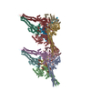
Structure visualization
Movie
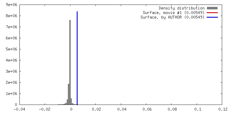
Surface view with section colored by density value
In the structure databanks used in Yorodumi, some data are registered as the other names, "COVID-19 virus" and "2019-nCoV". Here are the details of the virus and the list of structure data.
Jan 31, 2019. EMDB accession codes are about to change! (news from PDBe EMDB page)
EMDB accession codes are about to change! (news from PDBe EMDB page)
The allocation of 4 digits for EMDB accession codes will soon come to an end. Whilst these codes will remain in use, new EMDB accession codes will include an additional digit and will expand incrementally as the available range of codes is exhausted. The current 4-digit format prefixed with “EMD-” (i.e. EMD-XXXX) will advance to a 5-digit format (i.e. EMD-XXXXX), and so on. It is currently estimated that the 4-digit codes will be depleted around Spring 2019, at which point the 5-digit format will come into force.
The EM Navigator/Yorodumi systems omit the EMD- prefix.
Related info.:Q: What is EMD? / ID/Accession-code notation in Yorodumi/EM Navigator
Yorodumi is a browser for structure data from EMDB, PDB, SASBDB, etc.
This page is also the successor to EM Navigator detail page, and also detail information page/front-end page for Omokage search.
The word "yorodu" (or yorozu) is an old Japanese word meaning "ten thousand". "mi" (miru) is to see.
Related info.:EMDB / PDB / SASBDB / Comparison of 3 databanks / Yorodumi Search / Aug 31, 2016. New EM Navigator & Yorodumi / Yorodumi Papers / Jmol/JSmol / Function and homology information / Changes in new EM Navigator and Yorodumi
 Movie
Movie Controller


 Open data
 Basic information
 Map data
 Sample
 Influenza A virus
 Authors
 Citation


 Structure visualization
 Movie viewer
 UCSF Chimera
 Downloads & links
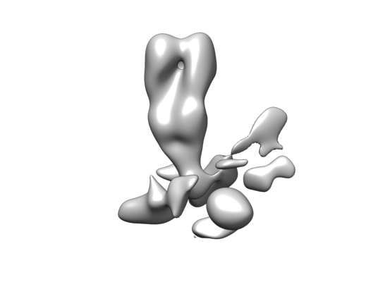 emd_9061.png
 http://ftp.pdbj.org/pub/emdb/structures/EMD-9061
 Z (Sec.)
 Y (Row.)
 X (Col.)


 Sample components
 Homo sapiens (human)
 Processing
 Electron microscopy