Movie
Movie Controller


+ Open data

- Basic information
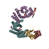
| Entry | Database: EMDB / ID: EMD-8927 | |||||||||
|---|---|---|---|---|---|---|---|---|---|---|
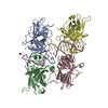
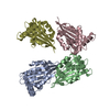
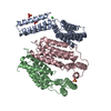
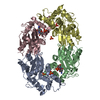
| Title | Wild-type p53-DNA assembly | |||||||||
 Map data Map data | Wild-type p53-DNA assembly | |||||||||
 Sample Sample |
| |||||||||
| Biological species |  Homo sapiens (human) / Human (human) Homo sapiens (human) / Human (human) | |||||||||
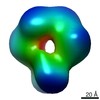
| Method | single particle reconstruction / negative staining / Resolution: 20.1 Å | |||||||||
 Authors Authors | Kelly DF / Dearnaley WJ / Liang Y | |||||||||
 Citation Citation | Journal: DNA Repair (Amst) / Year: 2019 Title: Correcting errors in the BRCA1 warning system. Authors: Yanping Liang / William J Dearnaley / Nick A Alden / Maria J Solares / Brian L Gilmore / Kevin J Pridham / A Cameron Varano / Zhi Sheng / Elizabeth Alli / Deborah F Kelly /  Abstract: Given its important role in human health and disease, remarkably little is known about the full-length three-dimensional (3D) molecular architecture of the breast cancer type 1 susceptibility protein ...Given its important role in human health and disease, remarkably little is known about the full-length three-dimensional (3D) molecular architecture of the breast cancer type 1 susceptibility protein (BRCA1), or its mechanisms to engage the tumor suppressor, TP53 (p53). Here, we show how a prevalent cancer-related mutation in the C-terminal region of the full-length protein, BRCA1, affects its structural properties, yet can be biochemically corrected to restore its functional capacity. As a downstream consequence of restoring the ubiquitin ligase activity of mutated BRCA1, the DNA repair response of p53 was enhanced in cellular extracts naturally deficient in BRCA1 protein expression. Complementary structural insights of p53 tetramers bound to DNA in different stage of the repair process support these biochemical findings in the context of human cancer cells. Equally important, we show how this knowledge can be used to lower the viability of breast cancer cells by modulating the stability of the BRCA1 protein and its associated players. | |||||||||
| History |
|
- Structure visualization
| Movie |
 Movie viewer Movie viewer |
|---|---|
| Structure viewer | EM map: SurfViewMolmilJmol/JSmol |
| Supplemental images |
 UCSF Chimera
- Downloads & links
-EMDB archive
| Map data | emd_8927.map.gz | 139.4 KB | EMDB map data format | |
|---|---|---|---|---|
| Header (meta data) | emd-8927-v30.xmlemd-8927.xml | 12.3 KB 12.3 KB | Display Display | EMDB header |
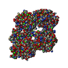
| Images | 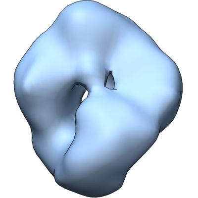 emd_8927.png emd_8927.png | 92.4 KB | ||
| Archive directory |  http://ftp.pdbj.org/pub/emdb/structures/EMD-8927ftp://ftp.pdbj.org/pub/emdb/structures/EMD-8927 http://ftp.pdbj.org/pub/emdb/structures/EMD-8927ftp://ftp.pdbj.org/pub/emdb/structures/EMD-8927 | HTTPS FTP |
-Related structure data


-Links
| EMDB pages | EMDB (EBI/PDBe) / EMDataResource |
|---|
-Map
| File | Download / File: emd_8927.map.gz / Format: CCP4 / Size: 3.8 MB / Type: IMAGE STORED AS FLOATING POINT NUMBER (4 BYTES) | ||||||||||||||||||||||||||||||||||||||||||||||||||||||||||||
|---|---|---|---|---|---|---|---|---|---|---|---|---|---|---|---|---|---|---|---|---|---|---|---|---|---|---|---|---|---|---|---|---|---|---|---|---|---|---|---|---|---|---|---|---|---|---|---|---|---|---|---|---|---|---|---|---|---|---|---|---|---|
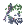
| Annotation | Wild-type p53-DNA assembly | ||||||||||||||||||||||||||||||||||||||||||||||||||||||||||||
| Projections & slices | Image control
Images are generated by Spider. | ||||||||||||||||||||||||||||||||||||||||||||||||||||||||||||
| Voxel size | X=Y=Z: 3.2 Å | ||||||||||||||||||||||||||||||||||||||||||||||||||||||||||||
| Density |
| ||||||||||||||||||||||||||||||||||||||||||||||||||||||||||||
| Symmetry | Space group: 1 | ||||||||||||||||||||||||||||||||||||||||||||||||||||||||||||
| Details | EMDB XML:
CCP4 map header:
| ||||||||||||||||||||||||||||||||||||||||||||||||||||||||||||
 Z (Sec.)
 Y (Row.)
 X (Col.)


-Supplemental data
- Sample components
-Entire : Wild-type p53-DNA assembly
| Entire | Name: Wild-type p53-DNA assembly |
|---|---|
| Components |
|
-Supramolecule #1: Wild-type p53-DNA assembly
| Supramolecule | Name: Wild-type p53-DNA assembly / type: complex / ID: 1 / Parent: 0 / Macromolecule list: all |
|---|---|
| Source (natural) | Organism: Homo sapiens (human) / Organ: Brain / Tissue: Brain |
| Molecular weight | Theoretical: 220 KDa |
-Macromolecule #1: Wild-type p53-DNA assembly
| Macromolecule | Name: Wild-type p53-DNA assembly / type: protein_or_peptide / ID: 1 / Enantiomer: DEXTRO |
|---|---|
| Source (natural) | Organism: Human (human) / Organ: Brain / Tissue: Brain |
| Sequence | String: MEEPQSDPSV EPPLSQETFS DLWKLLPENN VLSPLPSQAM DDLMLSPDDI EQWFTEDPGP DEAPRMPEAA PPVAPAPAAP TPAAPAPAPS WPLSSSVPSQ KTYQGSYGFR LGFLHSGTAK SVTCTYSPAL NKMFCQLAKT CPVQLWVDST PPPGTRVRAM AIYKQSQHMT ...String: MEEPQSDPSV EPPLSQETFS DLWKLLPENN VLSPLPSQAM DDLMLSPDDI EQWFTEDPGP DEAPRMPEAA PPVAPAPAAP TPAAPAPAPS WPLSSSVPSQ KTYQGSYGFR LGFLHSGTAK SVTCTYSPAL NKMFCQLAKT CPVQLWVDST PPPGTRVRAM AIYKQSQHMT EVVRRCPHHE RCSDSDGLAP PQHLIRVEGN LRVEYLDDRN TFRHSVVVPY EPPEVGSDCT TIHYNYMCNS SCMGGMNRRP ILTIITLEDS SGNLLGRNSF EVRVCACPGR DRRTEEENLR KKGEPHHELP PGSTKRALPN NTSSSPQPKK KPLDGEYFTL QIRGRERFEM FRELNEALEL KDAQAGKEPG GSRAHSSHLK SKKGQSTSRH KKLMFKTEGP DSD |
-Experimental details
-Structure determination
| Method | negative staining |
|---|---|
 Processing Processing | single particle reconstruction |
| Aggregation state | particle |
-Sample preparation
| Concentration | 0.02 mg/mL |
|---|---|
| Buffer | pH: 7.2 / Component - Name: HEPES buffer Details: 20 mM HEPES, pH 7.2, 150 mM NaCl, 10 mM CaCl2, 10 mM MgCl2 |
| Staining | Type: NEGATIVE / Material: Uranyl Formate / Details: 1% uranyl formate |
| Grid | Support film - Material: CARBON / Support film - topology: CONTINUOUS / Details: unspecified |
- Electron microscopy
| Microscope | FEI TECNAI SPIRIT |
|---|---|
| Image recording | Film or detector model: FEI EAGLE (2k x 2k) / Digitization - Sampling interval: 30.0 µm / Number grids imaged: 1 / Number real images: 20 / Average electron dose: 5.0 e/Å2 |
| Electron beam | Acceleration voltage: 120 kV / Electron source: LAB6 |
| Electron optics | Illumination mode: FLOOD BEAM / Imaging mode: BRIGHT FIELD / Cs: 2.0 mm / Nominal defocus min: -1.5 µm / Nominal magnification: 68000 |
| Sample stage | Specimen holder model: SIDE ENTRY, EUCENTRIC / Cooling holder cryogen: NITROGEN |
| Experimental equipment |  Model: Tecnai Spirit / Image courtesy: FEI Company |