 Movie
Movie Controller

[English] 日本語
 Yorodumi
- EMDB-8759: Yeast microtubule assembled using the slowly hydrolyzable analogu... -
+ Open data

- Basic information
| Entry | Database: EMDB / ID: EMD-8759 | ||||||||||||||||||||||||
|---|---|---|---|---|---|---|---|---|---|---|---|---|---|---|---|---|---|---|---|---|---|---|---|---|---|
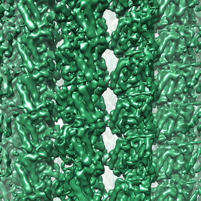
| Title | Yeast microtubule assembled using the slowly hydrolyzable analogue GTPgammaS | ||||||||||||||||||||||||
 Map data Map data | Yeast microtubule assembled using the slowly hydrolyzable analogue GTPgammaS | ||||||||||||||||||||||||
 Sample Sample |
| ||||||||||||||||||||||||
 Keywords Keywords | Cytoskeleton / tubulin / HYDROLASE | ||||||||||||||||||||||||
| Function / homology |  Function and homology information Function and homology informationnuclear migration by microtubule mediated pushing forces / nuclear division / mitotic spindle elongation / Platelet degranulation / homologous chromosome segregation / nuclear migration along microtubule / positive regulation of intracellular protein transport / tubulin complex / mitotic sister chromatid segregation / mitotic spindle assembly ...nuclear migration by microtubule mediated pushing forces / nuclear division / mitotic spindle elongation / Platelet degranulation / homologous chromosome segregation / nuclear migration along microtubule / positive regulation of intracellular protein transport / tubulin complex / mitotic sister chromatid segregation / mitotic spindle assembly / microtubule-based process / cytoplasmic microtubule organization / cytoskeleton organization / nuclear periphery / structural constituent of cytoskeleton / microtubule cytoskeleton organization / spindle / mitotic cell cycle / microtubule / Hydrolases; Acting on acid anhydrides; Acting on GTP to facilitate cellular and subcellular movement / response to antibiotic / hydrolase activity / GTPase activity / GTP binding / metal ion binding / nucleus / cytoplasm Similarity search - Function | ||||||||||||||||||||||||
| Biological species |  | ||||||||||||||||||||||||
| Method | helical reconstruction / cryo EM / Resolution: 6.0 Å | ||||||||||||||||||||||||
 Authors Authors | Howes SC / Geyer EA / LaFrance B / Zhang R / Kellogg EH / Westermann S / Rice LM / Nogales E | ||||||||||||||||||||||||
| Funding support |  United States, 7 items United States, 7 items
| ||||||||||||||||||||||||
 Citation Citation | Journal: J Cell Biol / Year: 2017 Title: Structural differences between yeast and mammalian microtubules revealed by cryo-EM. Authors: Stuart C Howes / Elisabeth A Geyer / Benjamin LaFrance / Rui Zhang / Elizabeth H Kellogg / Stefan Westermann / Luke M Rice / Eva Nogales /  Abstract: Microtubules are polymers of αβ-tubulin heterodimers essential for all eukaryotes. Despite sequence conservation, there are significant structural differences between microtubules assembled in ...Microtubules are polymers of αβ-tubulin heterodimers essential for all eukaryotes. Despite sequence conservation, there are significant structural differences between microtubules assembled in vitro from mammalian or budding yeast tubulin. Yeast MTs were not observed to undergo compaction at the interdimer interface as seen for mammalian microtubules upon GTP hydrolysis. Lack of compaction might reflect slower GTP hydrolysis or a different degree of allosteric coupling in the lattice. The microtubule plus end-tracking protein Bim1 binds yeast microtubules both between αβ-tubulin heterodimers, as seen for other organisms, and within tubulin dimers, but binds mammalian tubulin only at interdimer contacts. At the concentrations used in cryo-electron microscopy, Bim1 causes the compaction of yeast microtubules and induces their rapid disassembly. Our studies demonstrate structural differences between yeast and mammalian microtubules that likely underlie their differing polymerization dynamics. These differences may reflect adaptations to the demands of different cell size or range of physiological growth temperatures. | ||||||||||||||||||||||||
| History |
|
- Structure visualization
| Movie |
Movie viewer |
|---|---|
| Structure viewer | EM map: SurfViewMolmilJmol/JSmol |
| Supplemental images |
- Downloads & links
-EMDB archive
| Map data | emd_8759.map.gz | 481.9 MB | EMDB map data format | |
|---|---|---|---|---|
| Header (meta data) | emd-8759-v30.xmlemd-8759.xml | 13.3 KB 13.3 KB | Display Display | EMDB header |
| Images |  emd_8759.png emd_8759.png | 325.7 KB | ||
| Archive directory |  http://ftp.pdbj.org/pub/emdb/structures/EMD-8759ftp://ftp.pdbj.org/pub/emdb/structures/EMD-8759 http://ftp.pdbj.org/pub/emdb/structures/EMD-8759ftp://ftp.pdbj.org/pub/emdb/structures/EMD-8759 | HTTPS FTP |
-Related structure data
| Related structure data |  8755C  8756C  8757C  8758C  5w3fC  5w3hC  5w3jC C: citing same article ( |
|---|---|
| Similar structure data |

-Links
| EMDB pages | EMDB (EBI/PDBe) / EMDataResource |
|---|---|
| Related items in Molecule of the Month |


-Map
| File | Download / File: emd_8759.map.gz / Format: CCP4 / Size: 512 MB / Type: IMAGE STORED AS FLOATING POINT NUMBER (4 BYTES) | ||||||||||||||||||||||||||||||||||||||||||||||||||||||||||||||||||||
|---|---|---|---|---|---|---|---|---|---|---|---|---|---|---|---|---|---|---|---|---|---|---|---|---|---|---|---|---|---|---|---|---|---|---|---|---|---|---|---|---|---|---|---|---|---|---|---|---|---|---|---|---|---|---|---|---|---|---|---|---|---|---|---|---|---|---|---|---|---|
| Annotation | Yeast microtubule assembled using the slowly hydrolyzable analogue GTPgammaS | ||||||||||||||||||||||||||||||||||||||||||||||||||||||||||||||||||||
| Projections & slices | Image control
Images are generated by Spider. | ||||||||||||||||||||||||||||||||||||||||||||||||||||||||||||||||||||
| Voxel size | X=Y=Z: 1.32 Å | ||||||||||||||||||||||||||||||||||||||||||||||||||||||||||||||||||||
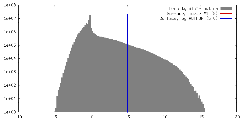
| Density |
| ||||||||||||||||||||||||||||||||||||||||||||||||||||||||||||||||||||
| Symmetry | Space group: 1 | ||||||||||||||||||||||||||||||||||||||||||||||||||||||||||||||||||||
| Details | EMDB XML:
CCP4 map header:
| ||||||||||||||||||||||||||||||||||||||||||||||||||||||||||||||||||||
 Z (Sec.)
 Y (Row.)
 X (Col.)


-Supplemental data
- Sample components
-Entire : Yeast microtubule assembled using the slowly hydrolyzable analogu...
| Entire | Name: Yeast microtubule assembled using the slowly hydrolyzable analogue GTPgammaS |
|---|---|
| Components |
|
-Supramolecule #1: Yeast microtubule assembled using the slowly hydrolyzable analogu...
| Supramolecule | Name: Yeast microtubule assembled using the slowly hydrolyzable analogue GTPgammaS type: complex / ID: 1 / Parent: 0 / Macromolecule list: all |
|---|---|
| Source (natural) | Organism: |
-Macromolecule #1: Tubulin alpha-1
| Macromolecule | Name: Tubulin alpha-1 / type: protein_or_peptide / ID: 1 / Enantiomer: LEVO |
|---|---|
| Source (natural) | Organism: |
| Sequence | String: MREVISINVG QAGCQIGNAC WELYSLEHGI KPDGHLEDGL SKPKGGEEGF STFFHETGYG KFVPRAIYV DLEPNVIDEV RNGPYKDLFH PEQLISGKED AANNYARGHY TVGREILGDV L DRIRKLAD QCDGLQGFLF THSLGGGTGS GLGSLLLEEL SAEYGKKSKL ...String: MREVISINVG QAGCQIGNAC WELYSLEHGI KPDGHLEDGL SKPKGGEEGF STFFHETGYG KFVPRAIYV DLEPNVIDEV RNGPYKDLFH PEQLISGKED AANNYARGHY TVGREILGDV L DRIRKLAD QCDGLQGFLF THSLGGGTGS GLGSLLLEEL SAEYGKKSKL EFAVYPAPQV ST SVVEPYN TVLTTHTTLE HADCTFMVDN EAIYDMCKRN LDIPRPSFAN LNNLIAQVVS SVT ASLRFD GSLNVDLNEF QTNLVPYPRI HFPLVSYSPV LSKSKAFHES NSVSEITNAC FEPG NQMVK CDPRDGKYMA TCLLYRGDVV TRDVQRAVEQ VKNKKTVQLV DWCPTGFKIG ICYEP PTAT PNSQLATVDR AVCMLSNTTS IAEAWKRIDR KFDLMYAKRA FVHWYVGEGM EEGEFT EAR EDLAALERDY IEVGADSYAE EEEF UniProtKB: Tubulin alpha-1 chain |
-Macromolecule #2: Tubulin beta
| Macromolecule | Name: Tubulin beta / type: protein_or_peptide / ID: 2 / Enantiomer: LEVO |
|---|---|
| Source (natural) | Organism: |
| Sequence | String: MREIIHISTG QCGNQIGAAF WETICGEHGL DFNGTYHGHD DIQKERLNVY FNEASSGKWV PRSINVDLE PGTIDAVRNS AIGNLFRPDN YIFGQSSAGN VWAKGHYTEG AELVDSVMDV I RREAEGCD SLQGFQITHS LGGGTGSGMG TLLISKIREE FPDRMMATFS ...String: MREIIHISTG QCGNQIGAAF WETICGEHGL DFNGTYHGHD DIQKERLNVY FNEASSGKWV PRSINVDLE PGTIDAVRNS AIGNLFRPDN YIFGQSSAGN VWAKGHYTEG AELVDSVMDV I RREAEGCD SLQGFQITHS LGGGTGSGMG TLLISKIREE FPDRMMATFS VLPSPKTSDT VV EPYNATL SVHQLVEHSD ETFCIDNEAL YDICQRTLKL NQPSYGDLNN LVSSVMSGVT TSL RYPGQL NSDLRKLAVN LVPFPRLHFF MVGYAPLTAI GSQSFRSLTV PELTQQMFDA KNMM AAADP RNGRYLTVAA FFRGKVSVKE VEDEMHKVQS KNSDYFVEWI PNNVQTAVCS VAPQG LDMA ATFIANSTSI QELFKRVGDQ FSAMFKRKAF LHWYTSEGMD ELEFSEAESN MNDLVS EYQ QYQEATVEDD EEVDENGDFG APQNQDEPIT ENFE UniProtKB: Tubulin beta chain |
-Experimental details
-Structure determination
| Method | cryo EM |
|---|---|
 Processing Processing | helical reconstruction |
| Aggregation state | helical array |
-Sample preparation
| Buffer | pH: 6.9 |
|---|---|
| Vitrification | Cryogen name: ETHANE / Chamber humidity: 100 % / Chamber temperature: 303 K / Instrument: FEI VITROBOT MARK IV |
- Electron microscopy
| Microscope | FEI TITAN |
|---|---|
| Image recording | Film or detector model: GATAN K2 SUMMIT (4k x 4k) / Detector mode: COUNTING / Average electron dose: 28.0 e/Å2 |
| Electron beam | Acceleration voltage: 300 kV / Electron source:  FIELD EMISSION GUN FIELD EMISSION GUN |
| Electron optics | Illumination mode: FLOOD BEAM / Imaging mode: BRIGHT FIELD |
-Image processing
| Final reconstruction | Applied symmetry - Helical parameters - Δz: 10.3 Å Applied symmetry - Helical parameters - Δ&Phi: -29.9 ° Applied symmetry - Helical parameters - Axial symmetry: C1 (asymmetric) Resolution.type: BY AUTHOR / Resolution: 6.0 Å / Resolution method: FSC 0.143 CUT-OFF / Software - Name: FREALIGN (ver. 9.11) / Number images used: 4999 |
|---|---|
| Startup model | Type of model: EMDB MAP |
| Final angle assignment | Type: NOT APPLICABLE |
