 Movie
Movie Controller

[English] 日本語
 Yorodumi
- EMDB-8757: Yeast microtubule stabilized with Taxol assembled from mutated tubulin -
+ Open data

- Basic information
| Entry | Database: EMDB / ID: EMD-8757 | ||||||||||||||||||||||||
|---|---|---|---|---|---|---|---|---|---|---|---|---|---|---|---|---|---|---|---|---|---|---|---|---|---|
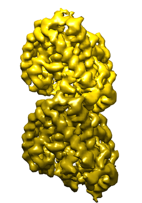
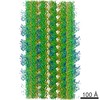
| Title | Yeast microtubule stabilized with Taxol assembled from mutated tubulin | ||||||||||||||||||||||||
 Map data Map data | Yeast microtubule stabilized with Taxol assembled from mutated tubulin | ||||||||||||||||||||||||
 Sample Sample |
| ||||||||||||||||||||||||
 Keywords Keywords | Cytoskeleton / tubulin / HYDROLASE | ||||||||||||||||||||||||
| Function / homology |  Function and homology information Function and homology informationnuclear migration by microtubule mediated pushing forces / nuclear division / mitotic spindle elongation / Platelet degranulation / homologous chromosome segregation / nuclear migration along microtubule / positive regulation of intracellular protein transport / tubulin complex / mitotic sister chromatid segregation / mitotic spindle assembly ...nuclear migration by microtubule mediated pushing forces / nuclear division / mitotic spindle elongation / Platelet degranulation / homologous chromosome segregation / nuclear migration along microtubule / positive regulation of intracellular protein transport / tubulin complex / mitotic sister chromatid segregation / mitotic spindle assembly / microtubule-based process / cytoplasmic microtubule organization / cytoskeleton organization / nuclear periphery / structural constituent of cytoskeleton / microtubule cytoskeleton organization / spindle / mitotic cell cycle / microtubule / Hydrolases; Acting on acid anhydrides; Acting on GTP to facilitate cellular and subcellular movement / response to antibiotic / hydrolase activity / GTPase activity / GTP binding / metal ion binding / nucleus / cytoplasm Similarity search - Function | ||||||||||||||||||||||||
| Biological species |  | ||||||||||||||||||||||||
| Method | helical reconstruction / cryo EM / Resolution: 4.0 Å | ||||||||||||||||||||||||
 Authors Authors | Howes SC / Geyer EA | ||||||||||||||||||||||||
| Funding support |  United States, 7 items United States, 7 items
| ||||||||||||||||||||||||
 Citation Citation | Journal: J Cell Biol / Year: 2017 Title: Structural differences between yeast and mammalian microtubules revealed by cryo-EM. Authors: Stuart C Howes / Elisabeth A Geyer / Benjamin LaFrance / Rui Zhang / Elizabeth H Kellogg / Stefan Westermann / Luke M Rice / Eva Nogales /  Abstract: Microtubules are polymers of αβ-tubulin heterodimers essential for all eukaryotes. Despite sequence conservation, there are significant structural differences between microtubules assembled in ...Microtubules are polymers of αβ-tubulin heterodimers essential for all eukaryotes. Despite sequence conservation, there are significant structural differences between microtubules assembled in vitro from mammalian or budding yeast tubulin. Yeast MTs were not observed to undergo compaction at the interdimer interface as seen for mammalian microtubules upon GTP hydrolysis. Lack of compaction might reflect slower GTP hydrolysis or a different degree of allosteric coupling in the lattice. The microtubule plus end-tracking protein Bim1 binds yeast microtubules both between αβ-tubulin heterodimers, as seen for other organisms, and within tubulin dimers, but binds mammalian tubulin only at interdimer contacts. At the concentrations used in cryo-electron microscopy, Bim1 causes the compaction of yeast microtubules and induces their rapid disassembly. Our studies demonstrate structural differences between yeast and mammalian microtubules that likely underlie their differing polymerization dynamics. These differences may reflect adaptations to the demands of different cell size or range of physiological growth temperatures. | ||||||||||||||||||||||||
| History |
|
- Structure visualization
| Movie |
Movie viewer |
|---|---|
| Structure viewer | EM map: SurfViewMolmilJmol/JSmol |
| Supplemental images |
- Downloads & links
-EMDB archive
| Map data | emd_8757.map.gz | 480.9 MB | EMDB map data format | |
|---|---|---|---|---|
| Header (meta data) | emd-8757-v30.xmlemd-8757.xml | 15.8 KB 15.8 KB | Display Display | EMDB header |
| Images |  emd_8757.png emd_8757.png | 107.2 KB | ||
| Filedesc metadata | emd-8757.cif.gz | 6 KB | ||
| Archive directory |  http://ftp.pdbj.org/pub/emdb/structures/EMD-8757ftp://ftp.pdbj.org/pub/emdb/structures/EMD-8757 http://ftp.pdbj.org/pub/emdb/structures/EMD-8757ftp://ftp.pdbj.org/pub/emdb/structures/EMD-8757 | HTTPS FTP |
-Related structure data
| Related structure data |  5w3jMC  8755C  8756C  8758C 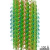 8759C  5w3fC  5w3hC C: citing same article ( M: atomic model generated by this map |
|---|---|
| Similar structure data |

-Links
| EMDB pages | EMDB (EBI/PDBe) / EMDataResource |
|---|---|
| Related items in Molecule of the Month |


-Map
| File | Download / File: emd_8757.map.gz / Format: CCP4 / Size: 512 MB / Type: IMAGE STORED AS FLOATING POINT NUMBER (4 BYTES) | ||||||||||||||||||||||||||||||||||||||||||||||||||||||||||||
|---|---|---|---|---|---|---|---|---|---|---|---|---|---|---|---|---|---|---|---|---|---|---|---|---|---|---|---|---|---|---|---|---|---|---|---|---|---|---|---|---|---|---|---|---|---|---|---|---|---|---|---|---|---|---|---|---|---|---|---|---|---|
| Annotation | Yeast microtubule stabilized with Taxol assembled from mutated tubulin | ||||||||||||||||||||||||||||||||||||||||||||||||||||||||||||
| Projections & slices | Image control
Images are generated by Spider. | ||||||||||||||||||||||||||||||||||||||||||||||||||||||||||||
| Voxel size | X=Y=Z: 1.32 Å | ||||||||||||||||||||||||||||||||||||||||||||||||||||||||||||
| Density |
| ||||||||||||||||||||||||||||||||||||||||||||||||||||||||||||
| Symmetry | Space group: 1 | ||||||||||||||||||||||||||||||||||||||||||||||||||||||||||||
| Details | EMDB XML:
CCP4 map header:
| ||||||||||||||||||||||||||||||||||||||||||||||||||||||||||||
 Z (Sec.)
 Y (Row.)
 X (Col.)


-Supplemental data
- Sample components
-Entire : Yeast microtubule stabilized with taxol assembled from mutated tubulin
| Entire | Name: Yeast microtubule stabilized with taxol assembled from mutated tubulin |
|---|---|
| Components |
|
-Supramolecule #1: Yeast microtubule stabilized with taxol assembled from mutated tubulin
| Supramolecule | Name: Yeast microtubule stabilized with taxol assembled from mutated tubulin type: complex / ID: 1 / Parent: 0 / Macromolecule list: #1-#2 |
|---|---|
| Source (natural) | Organism: |
-Macromolecule #1: Tubulin alpha-1 chain
| Macromolecule | Name: Tubulin alpha-1 chain / type: protein_or_peptide / ID: 1 / Number of copies: 1 / Enantiomer: LEVO |
|---|---|
| Source (natural) | Organism: |
| Molecular weight | Theoretical: 49.853867 KDa |
| Recombinant expression | Organism: |
| Sequence | String: MREVISINVG QAGCQIGNAC WELYSLEHGI KPDGHLEDGL SKPKGGEEGF STFFHETGYG KFVPRAIYVD LEPNVIDEVR NGPYKDLFH PEQLISGKED AANNYARGHY TVGREILGDV LDRIRKLADQ CDGLQGFLFT HSLGGGTGSG LGSLLLEELS A EYGKKSKL ...String: MREVISINVG QAGCQIGNAC WELYSLEHGI KPDGHLEDGL SKPKGGEEGF STFFHETGYG KFVPRAIYVD LEPNVIDEVR NGPYKDLFH PEQLISGKED AANNYARGHY TVGREILGDV LDRIRKLADQ CDGLQGFLFT HSLGGGTGSG LGSLLLEELS A EYGKKSKL EFAVYPAPQV STSVVEPYNT VLTTHTTLEH ADCTFMVDNE AIYDMCKRNL DIPRPSFANL NNLIAQVVSS VT ASLRFDG SLNVDLNEFQ TNLVPYPRIH FPLVSYSPVL SKSKAFHESN SVSEITNACF EPGNQMVKCD PRDGKYMATC LLY RGDVVT RDVQRAVEQV KNKKTVQLVD WCPTGFKIGI CYEPPTATPN SQLATVDRAV CMLSNTTSIA EAWKRIDRKF DLMY AKRAF VHWYVGEGME EGEFTEARED LAALERDYIE VGADSYAEEE EF UniProtKB: Tubulin alpha-1 chain |
-Macromolecule #2: Tubulin beta chain
| Macromolecule | Name: Tubulin beta chain / type: protein_or_peptide / ID: 2 / Number of copies: 1 / Enantiomer: LEVO |
|---|---|
| Source (natural) | Organism: |
| Molecular weight | Theoretical: 51.089668 KDa |
| Recombinant expression | Organism: |
| Sequence | String: MREIIHISTG QCGNQIGAKF WEVICDEHGL DFNGTYHGHD DIQKERLNVY FNEASSGKWV PRSINVDLEP GTIDAVRNSA IGNLFRPDN YIFGQSSAGN VWAKGHYTEG AELVDSVMDV IRREAEGCDS LQGFQITHSL GGGTGSGMGT LLISKIREEF P DRMMATFS ...String: MREIIHISTG QCGNQIGAKF WEVICDEHGL DFNGTYHGHD DIQKERLNVY FNEASSGKWV PRSINVDLEP GTIDAVRNSA IGNLFRPDN YIFGQSSAGN VWAKGHYTEG AELVDSVMDV IRREAEGCDS LQGFQITHSL GGGTGSGMGT LLISKIREEF P DRMMATFS VLPSPKTSDT VVEPYNATLS VHQLVEHSDE TFCIDNEALY DICQRTLKLN QPSYGDLNHL VSSVMSGVTT SL RYPGQLN SDLRKLAVNL VPFPRLHFFM VGFAPLTAIG SQSFRSLTVP ELTQQMFDAK NMMAAADPRN GRYLTVAAFF RGK VSVKEV EDEMHKVQSK NSDYFVEWIP NNVQTAVCSV APQGLDMAAT FIANSTSIQE LFKRVGDQFS AMFKRKAFLH WYTS EGMDE LEFSEAESNM NDLVSEYQQY QEATVEDDEE VDENGDFGAP QNQDEPITEN FE UniProtKB: Tubulin beta chain |
-Macromolecule #3: GUANOSINE-5'-TRIPHOSPHATE
| Macromolecule | Name: GUANOSINE-5'-TRIPHOSPHATE / type: ligand / ID: 3 / Number of copies: 1 / Formula: GTP |
|---|---|
| Molecular weight | Theoretical: 523.18 Da |
| Chemical component information |  ChemComp-GTP: |
-Macromolecule #4: MAGNESIUM ION
| Macromolecule | Name: MAGNESIUM ION / type: ligand / ID: 4 / Number of copies: 1 / Formula: MG |
|---|---|
| Molecular weight | Theoretical: 24.305 Da |
-Macromolecule #5: GUANOSINE-5'-DIPHOSPHATE
| Macromolecule | Name: GUANOSINE-5'-DIPHOSPHATE / type: ligand / ID: 5 / Number of copies: 1 / Formula: GDP |
|---|---|
| Molecular weight | Theoretical: 443.201 Da |
| Chemical component information |  ChemComp-GDP: |
-Macromolecule #6: TAXOL
| Macromolecule | Name: TAXOL / type: ligand / ID: 6 / Number of copies: 1 / Formula: TA1 |
|---|---|
| Molecular weight | Theoretical: 853.906 Da |
| Chemical component information |  ChemComp-TA1: |
-Experimental details
-Structure determination
| Method | cryo EM |
|---|---|
 Processing Processing | helical reconstruction |
| Aggregation state | helical array |
-Sample preparation
| Buffer | pH: 6.9 |
|---|---|
| Vitrification | Cryogen name: ETHANE / Chamber humidity: 100 % / Chamber temperature: 303 K / Instrument: FEI VITROBOT MARK IV |
- Electron microscopy
| Microscope | FEI TITAN |
|---|---|
| Image recording | Film or detector model: GATAN K2 SUMMIT (4k x 4k) / Detector mode: COUNTING / Average electron dose: 28.0 e/Å2 |
| Electron beam | Acceleration voltage: 300 kV / Electron source:  FIELD EMISSION GUN FIELD EMISSION GUN |
| Electron optics | Illumination mode: FLOOD BEAM / Imaging mode: BRIGHT FIELD |
| Sample stage | Specimen holder model: GATAN 626 SINGLE TILT LIQUID NITROGEN CRYO TRANSFER HOLDER Cooling holder cryogen: NITROGEN |
-Image processing
| Final reconstruction | Applied symmetry - Helical parameters - Δz: 10.41 Å Applied symmetry - Helical parameters - Δ&Phi: -29.87 ° Applied symmetry - Helical parameters - Axial symmetry: C1 (asymmetric) Algorithm: FOURIER SPACE / Resolution.type: BY AUTHOR / Resolution: 4.0 Å / Resolution method: FSC 0.143 CUT-OFF / Software - Name: FREALIGN (ver. 9.11) / Number images used: 30101 |
|---|---|
| Startup model | Type of model: EMDB MAP |
| Final angle assignment | Type: NOT APPLICABLE |
