 ムービー
ムービー コントローラー


+ データを開く

- 基本情報
基本情報
| 登録情報 | データベース: EMDB / ID: EMD-8589 | |||||||||
|---|---|---|---|---|---|---|---|---|---|---|
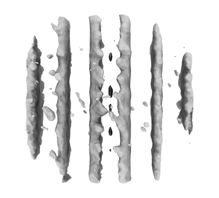
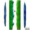
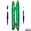
| タイトル | Subtomogram average of microtubules from Trypanosoma brucei | |||||||||
 マップデータ マップデータ | Subtomogram average of microtubules from Trypanosoma brucei | |||||||||
 試料 試料 |
| |||||||||
| 生物種 |  | |||||||||
| 手法 | サブトモグラム平均法 / クライオ電子顕微鏡法 / 解像度: 55.0 Å | |||||||||
 データ登録者 データ登録者 | Chen M / Sun SY / Ludtke SJ / He CY / Schmid MF / Chiu W | |||||||||
| 資金援助 |  米国, 1件 米国, 1件
| |||||||||
 引用 引用 | ジャーナル: Nat Methods / 年: 2017 タイトル: Convolutional neural networks for automated annotation of cellular cryo-electron tomograms. 著者: Muyuan Chen / Wei Dai / Stella Y Sun / Darius Jonasch / Cynthia Y He / Michael F Schmid / Wah Chiu / Steven J Ludtke /  要旨: Cellular electron cryotomography offers researchers the ability to observe macromolecules frozen in action in situ, but a primary challenge with this technique is identifying molecular components ...Cellular electron cryotomography offers researchers the ability to observe macromolecules frozen in action in situ, but a primary challenge with this technique is identifying molecular components within the crowded cellular environment. We introduce a method that uses neural networks to dramatically reduce the time and human effort required for subcellular annotation and feature extraction. Subsequent subtomogram classification and averaging yield in situ structures of molecular components of interest. The method is available in the EMAN2.2 software package. | |||||||||
| 履歴 |
|
- 構造の表示
構造の表示
| ムービー |
 ムービービューア ムービービューア |
|---|---|
| 構造ビューア | EMマップ: SurfViewMolmilJmol/JSmol |
| 添付画像 |
- ダウンロードとリンク
-EMDBアーカイブ
| マップデータ | emd_8589.map.gz | 25 MB | EMDBマップデータ形式 | |
|---|---|---|---|---|
| ヘッダ (付随情報) | emd-8589-v30.xmlemd-8589.xml | 9.1 KB 9.1 KB | 表示 表示 | EMDBヘッダ |
| 画像 |  emd_8589.png emd_8589.png | 52.9 KB | ||
| アーカイブディレクトリ |  http://ftp.pdbj.org/pub/emdb/structures/EMD-8589ftp://ftp.pdbj.org/pub/emdb/structures/EMD-8589 http://ftp.pdbj.org/pub/emdb/structures/EMD-8589ftp://ftp.pdbj.org/pub/emdb/structures/EMD-8589 | HTTPS FTP |
-検証レポート
| 文書・要旨 | emd_8589_validation.pdf.gz | 78.2 KB | 表示 | EMDB検証レポート |
|---|---|---|---|---|
| 文書・詳細版 | emd_8589_full_validation.pdf.gz | 77.2 KB | 表示 | |
| XML形式データ | emd_8589_validation.xml.gz | 495 B | 表示 | |
| アーカイブディレクトリ | https://ftp.pdbj.org/pub/emdb/validation_reports/EMD-8589ftp://ftp.pdbj.org/pub/emdb/validation_reports/EMD-8589 | HTTPS FTP |
-関連構造データ
-リンク
| EMDBのページ | EMDB (EBI/PDBe) / EMDataResource |
|---|---|
| 「今月の分子」の関連する項目 |

-マップ
| ファイル | ダウンロード / ファイル: emd_8589.map.gz / 形式: CCP4 / 大きさ: 27 MB / タイプ: IMAGE STORED AS FLOATING POINT NUMBER (4 BYTES) | ||||||||||||||||||||||||||||||||||||||||||||||||||||||||||||||||||||
|---|---|---|---|---|---|---|---|---|---|---|---|---|---|---|---|---|---|---|---|---|---|---|---|---|---|---|---|---|---|---|---|---|---|---|---|---|---|---|---|---|---|---|---|---|---|---|---|---|---|---|---|---|---|---|---|---|---|---|---|---|---|---|---|---|---|---|---|---|---|
| 注釈 | Subtomogram average of microtubules from Trypanosoma brucei | ||||||||||||||||||||||||||||||||||||||||||||||||||||||||||||||||||||
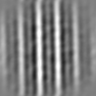
| 投影像・断面図 | 画像のコントロール
画像は Spider により作成 | ||||||||||||||||||||||||||||||||||||||||||||||||||||||||||||||||||||
| ボクセルのサイズ |
| ||||||||||||||||||||||||||||||||||||||||||||||||||||||||||||||||||||
| 密度 |
| ||||||||||||||||||||||||||||||||||||||||||||||||||||||||||||||||||||
| 対称性 | 空間群: 1 | ||||||||||||||||||||||||||||||||||||||||||||||||||||||||||||||||||||
| 詳細 | EMDB XML:
CCP4マップ ヘッダ情報:
| ||||||||||||||||||||||||||||||||||||||||||||||||||||||||||||||||||||
 Z (Sec.)
 Y (Row.)
 X (Col.)


-添付データ
- 試料の構成要素
-全体 : Microtubule
| 全体 | 名称: Microtubule |
|---|---|
| 要素 |
|
-超分子 #1: Microtubule
| 超分子 | 名称: Microtubule / タイプ: complex / ID: 1 / 親要素: 0 |
|---|---|
| 由来(天然) | 生物種: |
-実験情報
-構造解析
| 手法 | クライオ電子顕微鏡法 |
|---|---|
 解析 解析 | サブトモグラム平均法 |
| 試料の集合状態 | cell |
-試料調製
| 緩衝液 | pH: 7.2 |
|---|---|
| 凍結 | 凍結剤: ETHANE |
- 電子顕微鏡法
| 顕微鏡 | JEOL 2200FS |
|---|---|
| 撮影 | フィルム・検出器のモデル: DIRECT ELECTRON DE-12 (4k x 3k) 検出モード: INTEGRATING / 平均電子線量: 2.0 e/Å2 |
| 電子線 | 加速電圧: 200 kV / 電子線源:  FIELD EMISSION GUN FIELD EMISSION GUN |
| 電子光学系 | 照射モード: FLOOD BEAM / 撮影モード: BRIGHT FIELD |
-画像解析
| 最終 再構成 | 想定した対称性 - 点群: C1 (非対称) / アルゴリズム: BACK PROJECTION / 解像度のタイプ: BY AUTHOR / 解像度: 55.0 Å / 解像度の算出法: FSC 0.143 CUT-OFF / ソフトウェア - 名称: EMAN (ver. 2.2) / 使用したサブトモグラム数: 511 |
|---|---|
| 抽出 | トモグラム数: 1 / 使用した粒子像数: 511 / 参照モデル: Reference free averaging / 手法: Automated tomogram segmentation / ソフトウェア - 名称: EMAN (ver. 2.2) / ソフトウェア - 詳細: Tomoseg tool |
| 最終 角度割当 | タイプ: OTHER / ソフトウェア - 名称: EMAN (ver. 2.2) / 詳細: Single particle tomography tool in EMAN2 |
