 Movie
Movie Controller

[English] 日本語
 Yorodumi
- EMDB-10460: Sub-tomogram average of the Sulfolobus acidocaldarius S-layer in situ -
+ Open data

- Basic information
| Entry | Database: EMDB / ID: EMD-10460 | |||||||||
|---|---|---|---|---|---|---|---|---|---|---|
| Title | Sub-tomogram average of the Sulfolobus acidocaldarius S-layer in situ | |||||||||
 Map data Map data | Sub-tomogram averaging of the Sulfolobus acidocaldarius S-layer. | |||||||||
 Sample Sample |
| |||||||||
| Biological species |   Sulfolobus islandicus (archaea) Sulfolobus islandicus (archaea) | |||||||||
| Method | subtomogram averaging / cryo EM / Resolution: 56.0 Å | |||||||||
 Authors Authors | Daum B / Gambelli L | |||||||||
| Funding support |  United Kingdom, 1 items United Kingdom, 1 items
| |||||||||
 Citation Citation | Journal: Proc Natl Acad Sci U S A / Year: 2019 Title: Architecture and modular assembly of S-layers revealed by electron cryotomography. Authors: Lavinia Gambelli / Benjamin H Meyer / Mathew McLaren / Kelly Sanders / Tessa E F Quax / Vicki A M Gold / Sonja-Verena Albers / Bertram Daum /  Abstract: Surface protein layers (S-layers) often form the only structural component of the archaeal cell wall and are therefore important for cell survival. S-layers have a plethora of cellular functions ...Surface protein layers (S-layers) often form the only structural component of the archaeal cell wall and are therefore important for cell survival. S-layers have a plethora of cellular functions including maintenance of cell shape, osmotic, and mechanical stability, the formation of a semipermeable protective barrier around the cell, and cell-cell interaction, as well as surface adhesion. Despite the central importance of S-layers for archaeal life, their 3-dimensional (3D) architecture is still poorly understood. Here we present detailed 3D electron cryomicroscopy maps of archaeal S-layers from 3 different strains. We were able to pinpoint the positions and determine the structure of the 2 subunits SlaA and SlaB. We also present a model describing the assembly of the mature S-layer. #1: Journal: To Be PublishedTitle: Architecture and modular assembly of Sulfolobus S-layers revealed by electron cryo-tomography Authors: Gambelli L / Meyer BH / McLaren M / Sanders K / Quax TEF / Gold VAM / Albers SVA / Daum B | |||||||||
| History |
|
- Structure visualization
| Movie |
 Movie viewer Movie viewer |
|---|---|
| Structure viewer | EM map: SurfViewMolmilJmol/JSmol |
| Supplemental images |

- Downloads & links
-EMDB archive
| Map data | emd_10460.map.gz | 984.3 KB | EMDB map data format | |
|---|---|---|---|---|
| Header (meta data) | emd-10460-v30.xmlemd-10460.xml | 9.9 KB 9.9 KB | Display Display | EMDB header |
| Images | 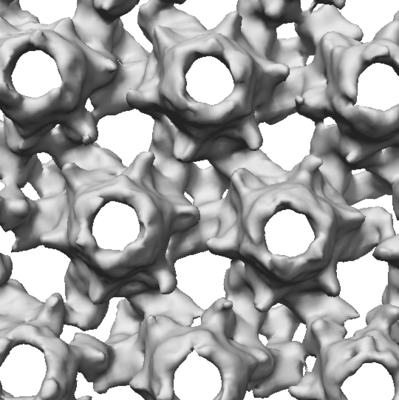 emd_10460.png emd_10460.png | 112.2 KB | ||
| Archive directory |  http://ftp.pdbj.org/pub/emdb/structures/EMD-10460ftp://ftp.pdbj.org/pub/emdb/structures/EMD-10460 http://ftp.pdbj.org/pub/emdb/structures/EMD-10460ftp://ftp.pdbj.org/pub/emdb/structures/EMD-10460 | HTTPS FTP |
-Related structure data
| Related structure data | C: citing same article ( |
|---|---|
| Similar structure data |


-Links
| EMDB pages | EMDB (EBI/PDBe) / EMDataResource |
|---|
-Map
| File | Download / File: emd_10460.map.gz / Format: CCP4 / Size: 1.5 MB / Type: IMAGE STORED AS FLOATING POINT NUMBER (4 BYTES) | ||||||||||||||||||||||||||||||||||||||||||||||||||||||||||||||||||||
|---|---|---|---|---|---|---|---|---|---|---|---|---|---|---|---|---|---|---|---|---|---|---|---|---|---|---|---|---|---|---|---|---|---|---|---|---|---|---|---|---|---|---|---|---|---|---|---|---|---|---|---|---|---|---|---|---|---|---|---|---|---|---|---|---|---|---|---|---|---|
| Annotation | Sub-tomogram averaging of the Sulfolobus acidocaldarius S-layer. | ||||||||||||||||||||||||||||||||||||||||||||||||||||||||||||||||||||
| Projections & slices | Image control
Images are generated by Spider. generated in cubic-lattice coordinate | ||||||||||||||||||||||||||||||||||||||||||||||||||||||||||||||||||||
| Voxel size | X=Y=Z: 10.73 Å | ||||||||||||||||||||||||||||||||||||||||||||||||||||||||||||||||||||
| Density |
| ||||||||||||||||||||||||||||||||||||||||||||||||||||||||||||||||||||
| Symmetry | Space group: 1 | ||||||||||||||||||||||||||||||||||||||||||||||||||||||||||||||||||||
| Details | EMDB XML:
CCP4 map header:
| ||||||||||||||||||||||||||||||||||||||||||||||||||||||||||||||||||||
 Z (Sec.)
 Y (Row.)
 X (Col.)


-Supplemental data
- Sample components
-Entire : SlaA/SlaB array
| Entire | Name: SlaA/SlaB array |
|---|---|
| Components |
|
-Supramolecule #1: SlaA/SlaB array
| Supramolecule | Name: SlaA/SlaB array / type: complex / ID: 1 / Parent: 0 |
|---|---|
| Source (natural) | Organism: Sulfolobus islandicus (archaea) |
-Experimental details
-Structure determination
| Method | cryo EM |
|---|---|
 Processing Processing | subtomogram averaging |
| Aggregation state | 2D array |
-Sample preparation
| Buffer | pH: 3 |
|---|---|
| Vitrification | Cryogen name: ETHANE |
- Electron microscopy
| Microscope | FEI POLARA 300 |
|---|---|
| Specialist optics | Energy filter - Slit width: 40 eV |
| Image recording | #0 - Image recording ID: 1 / #0 - Film or detector model: GATAN ULTRASCAN 4000 (4k x 4k) / #0 - Detector mode: COUNTING / #0 - Average electron dose: 1.8 e/Å2 / #1 - Image recording ID: 2 / #1 - Film or detector model: GATAN ULTRASCAN 4000 (4k x 4k) / #1 - Average electron dose: 1.8 e/Å2 |
| Electron beam | Acceleration voltage: 300 kV / Electron source:  FIELD EMISSION GUN FIELD EMISSION GUN |
| Electron optics | Illumination mode: FLOOD BEAM / Imaging mode: BRIGHT FIELD |
| Sample stage | Cooling holder cryogen: NITROGEN |
| Experimental equipment |  Model: Tecnai Polara / Image courtesy: FEI Company |
