 Movie
Movie Controller

[English] 日本語
 Yorodumi
- EMDB-8558: Negative stain reconstruction of Cas1-Cas2/3 complex of type I-F ... -
+ Open data

- Basic information
| Entry | Database: EMDB / ID: EMD-8558 | |||||||||
|---|---|---|---|---|---|---|---|---|---|---|
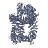
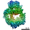
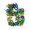
| Title | Negative stain reconstruction of Cas1-Cas2/3 complex of type I-F CRISPR system from Pseudomonas aeruginosa (PA14) | |||||||||
 Map data Map data | Negative stain reconstruction of Pseudomonas aeruginosa (PA14) Cas1-Cas2/3 complex | |||||||||
 Sample Sample |
| |||||||||
| Function / homology |  Function and homology information Function and homology informationmaintenance of CRISPR repeat elements / helicase activity / DNA endonuclease activity / Hydrolases; Acting on acid anhydrides; Acting on acid anhydrides to facilitate cellular and subcellular movement / defense response to virus / Hydrolases; Acting on ester bonds / hydrolase activity / DNA binding / ATP binding / metal ion binding / identical protein binding Similarity search - Function | |||||||||
| Biological species |  Pseudomonas aeruginosa PA14 (bacteria) Pseudomonas aeruginosa PA14 (bacteria) | |||||||||
| Method | single particle reconstruction / negative staining / Resolution: 15.6 Å | |||||||||
 Authors Authors | Chowdhury S / Lander GC / Rollins MF / Carter J / Golden MS / Wilkinson RA / Bondy-Denomy J / Wiedenheft B | |||||||||
 Citation Citation | Journal: Proc Natl Acad Sci U S A / Year: 2017 Title: Cas1 and the Csy complex are opposing regulators of Cas2/3 nuclease activity. Authors: MaryClare F Rollins / Saikat Chowdhury / Joshua Carter / Sarah M Golden / Royce A Wilkinson / Joseph Bondy-Denomy / Gabriel C Lander / Blake Wiedenheft /  Abstract: The type I-F CRISPR adaptive immune system in (PA14) consists of two CRISPR loci and six CRISPR-associated () genes. Type I-F systems rely on a CRISPR RNA (crRNA)-guided surveillance complex (Csy ...The type I-F CRISPR adaptive immune system in (PA14) consists of two CRISPR loci and six CRISPR-associated () genes. Type I-F systems rely on a CRISPR RNA (crRNA)-guided surveillance complex (Csy complex) to bind foreign DNA and recruit a -acting nuclease (i.e., Cas2/3) for target degradation. In most type I systems, Cas2 and Cas3 are separate proteins involved in adaptation and interference, respectively. However, in I-F systems, these proteins are fused into a single polypeptide. Here we use biochemical and structural methods to show that two molecules of Cas2/3 assemble with four molecules of Cas1 (Cas2/3:Cas1) into a four-lobed propeller-shaped structure, where the two Cas2 domains form a central hub (twofold axis of symmetry) flanked by two Cas1 lobes and two Cas3 lobes. We show that the Cas1 subunits repress Cas2/3 nuclease activity and that foreign DNA recognition by the Csy complex activates Cas2/3, resulting in bidirectional degradation of DNA targets. Collectively, this work provides a structure of the Cas1-2/3 complex and explains how Cas1 and the target-bound Csy complex play opposing roles in the regulation of Cas2/3 nuclease activity. | |||||||||
| History |
|
- Structure visualization
| Movie |
Movie viewer |
|---|---|
| Structure viewer | EM map: SurfViewMolmilJmol/JSmol |
| Supplemental images |
 UCSF Chimera
- Downloads & links
-EMDB archive
| Map data | emd_8558.map.gz | 2.4 MB | EMDB map data format | |
|---|---|---|---|---|
| Header (meta data) | emd-8558-v30.xmlemd-8558.xml | 13.5 KB 13.5 KB | Display Display | EMDB header |
| Images | 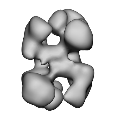 emd_8558.png emd_8558.png | 46.8 KB | ||
| Archive directory |  http://ftp.pdbj.org/pub/emdb/structures/EMD-8558ftp://ftp.pdbj.org/pub/emdb/structures/EMD-8558 http://ftp.pdbj.org/pub/emdb/structures/EMD-8558ftp://ftp.pdbj.org/pub/emdb/structures/EMD-8558 | HTTPS FTP |
-Related structure data
| Similar structure data |
|---|


-Links
| EMDB pages | EMDB (EBI/PDBe) / EMDataResource |
|---|---|
| Related items in Molecule of the Month |

-Map
| File | Download / File: emd_8558.map.gz / Format: CCP4 / Size: 3.4 MB / Type: IMAGE STORED AS FLOATING POINT NUMBER (4 BYTES) | ||||||||||||||||||||||||||||||||||||||||||||||||||||||||||||||||||||
|---|---|---|---|---|---|---|---|---|---|---|---|---|---|---|---|---|---|---|---|---|---|---|---|---|---|---|---|---|---|---|---|---|---|---|---|---|---|---|---|---|---|---|---|---|---|---|---|---|---|---|---|---|---|---|---|---|---|---|---|---|---|---|---|---|---|---|---|---|---|
| Annotation | Negative stain reconstruction of Pseudomonas aeruginosa (PA14) Cas1-Cas2/3 complex | ||||||||||||||||||||||||||||||||||||||||||||||||||||||||||||||||||||
| Projections & slices | Image control
Images are generated by Spider. | ||||||||||||||||||||||||||||||||||||||||||||||||||||||||||||||||||||
| Voxel size | X=Y=Z: 4.1 Å | ||||||||||||||||||||||||||||||||||||||||||||||||||||||||||||||||||||
| Density |
| ||||||||||||||||||||||||||||||||||||||||||||||||||||||||||||||||||||
| Symmetry | Space group: 1 | ||||||||||||||||||||||||||||||||||||||||||||||||||||||||||||||||||||
| Details | EMDB XML:
CCP4 map header:
| ||||||||||||||||||||||||||||||||||||||||||||||||||||||||||||||||||||
 Z (Sec.)
 Y (Row.)
 X (Col.)


-Supplemental data
- Sample components
-Entire : Complex containing dimer of Cas2/3 protein and tetramer of Cas1 p...
| Entire | Name: Complex containing dimer of Cas2/3 protein and tetramer of Cas1 protein. |
|---|---|
| Components |
|
-Supramolecule #1: Complex containing dimer of Cas2/3 protein and tetramer of Cas1 p...
| Supramolecule | Name: Complex containing dimer of Cas2/3 protein and tetramer of Cas1 protein. type: complex / ID: 1 / Parent: 0 |
|---|---|
| Source (natural) | Organism: Pseudomonas aeruginosa PA14 (bacteria) |
| Recombinant expression | Organism: |
| Molecular weight | Theoretical: 386 KDa |
-Experimental details
-Structure determination
| Method | negative staining |
|---|---|
 Processing Processing | single particle reconstruction |
| Aggregation state | particle |
-Sample preparation
| Concentration | 0.03 mg/mL |
|---|---|
| Buffer | pH: 7.5 / Details: 20 mM HEPES, 150 mM KCl |
| Staining | Type: NEGATIVE / Material: Uranyl Formate Details: Sample absorbed on carbon surface was stained with 2% (w/v) Uranyl Formate solution, and finally air-dried after blotting off excess stain. |
| Grid | Model: 400 mesh Cu-Rh maxtaform grids (Electron Microscopy Sciences) that were coated with a thin layer of amorphous carbon. Material: COPPER/RHODIUM / Mesh: 400 / Support film - Material: CARBON / Support film - topology: CONTINUOUS / Pretreatment - Type: GLOW DISCHARGE / Pretreatment - Atmosphere: AIR / Pretreatment - Pressure: 0.02 kPa / Details: 15mA current |
| Details | Mono dispersed protein solution |
- Electron microscopy
| Microscope | FEI TECNAI SPIRIT |
|---|---|
| Temperature | Min: 293.0 K / Max: 295.0 K |
| Image recording | Film or detector model: TVIPS TEMCAM-F416 (4k x 4k) / Digitization - Dimensions - Width: 4096 pixel / Digitization - Dimensions - Height: 4096 pixel / Digitization - Sampling interval: 15.6 µm / Number grids imaged: 1 / Number real images: 1159 / Average exposure time: 0.479 sec. / Average electron dose: 30.0 e/Å2 |
| Electron beam | Acceleration voltage: 120 kV / Electron source: LAB6 |
| Electron optics | C2 aperture diameter: 100.0 µm / Calibrated defocus max: 0.99 µm / Calibrated defocus min: 0.99 µm / Illumination mode: FLOOD BEAM / Imaging mode: BRIGHT FIELD / Cs: 2.7 mm / Nominal defocus max: 1.0 µm / Nominal defocus min: 1.0 µm / Nominal magnification: 52000 |
| Sample stage | Specimen holder model: SIDE ENTRY, EUCENTRIC / Cooling holder cryogen: NITROGEN |
| Experimental equipment |  Model: Tecnai Spirit / Image courtesy: FEI Company |
