 Movie
Movie Controller


+ Open data

- Basic information
| Entry | Database: EMDB / ID: EMD-8538 | |||||||||
|---|---|---|---|---|---|---|---|---|---|---|
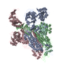
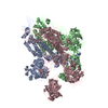
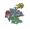
| Title | Structure of the p53/human RNA polymerase II assembly | |||||||||
 Map data Map data | The cryo-EM 3D reconstruction of p53-bound human RNA Polymerase II | |||||||||
 Sample Sample |
| |||||||||
| Biological species |  Homo sapiens (human) Homo sapiens (human) | |||||||||
| Method | single particle reconstruction / cryo EM / Resolution: 10.8 Å | |||||||||
 Authors Authors | Liu WL / Singh SK | |||||||||
 Citation Citation | Journal: Genes Dev / Year: 2016 Title: Structural visualization of the p53/RNA polymerase II assembly. Authors: Sameer K Singh / Zhen Qiao / Lihua Song / Vijay Jani / William Rice / Edward Eng / Robert A Coleman / Wei-Li Liu /  Abstract: The master tumor suppressor p53 activates transcription in response to various cellular stresses in part by facilitating recruitment of the transcription machinery to DNA. Recent studies have ...The master tumor suppressor p53 activates transcription in response to various cellular stresses in part by facilitating recruitment of the transcription machinery to DNA. Recent studies have documented a direct yet poorly characterized interaction between p53 and RNA polymerase II (Pol II). Therefore, we dissected the human p53/Pol II interaction via single-particle cryo-electron microscopy, structural docking, and biochemical analyses. This study reveals that p53 binds Pol II via the Rpb1 and Rpb2 subunits, bridging the DNA-binding cleft of Pol II proximal to the upstream DNA entry site. In addition, the key DNA-binding surface of p53, frequently disrupted in various cancers, remains exposed within the assembly. Furthermore, the p53/Pol II cocomplex displays a closed conformation as defined by the position of the Pol II clamp domain. Notably, the interaction of p53 and Pol II leads to increased Pol II elongation activity. These findings indicate that p53 may structurally regulate DNA-binding functions of Pol II via the clamp domain, thereby providing insights into p53-regulated Pol II transcription. | |||||||||
| History |
|
- Structure visualization
| Movie |
 Movie viewer Movie viewer |
|---|---|
| Structure viewer | EM map: SurfViewMolmilJmol/JSmol |
| Supplemental images |

- Downloads & links
-EMDB archive
| Map data | emd_8538.map.gz | 96.7 MB | EMDB map data format | |
|---|---|---|---|---|
| Header (meta data) | emd-8538-v30.xmlemd-8538.xml | 12.5 KB 12.5 KB | Display Display | EMDB header |
| Images | 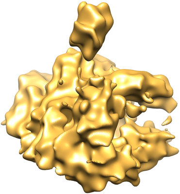 emd_8538.png emd_8538.png | 200.6 KB | ||
| Archive directory |  http://ftp.pdbj.org/pub/emdb/structures/EMD-8538ftp://ftp.pdbj.org/pub/emdb/structures/EMD-8538 http://ftp.pdbj.org/pub/emdb/structures/EMD-8538ftp://ftp.pdbj.org/pub/emdb/structures/EMD-8538 | HTTPS FTP |
-Related structure data
| Similar structure data |
|---|


-Links
| EMDB pages | EMDB (EBI/PDBe) / EMDataResource |
|---|
-Map
| File | Download / File: emd_8538.map.gz / Format: CCP4 / Size: 103 MB / Type: IMAGE STORED AS FLOATING POINT NUMBER (4 BYTES) | ||||||||||||||||||||||||||||||||||||||||||||||||||||||||||||||||||||
|---|---|---|---|---|---|---|---|---|---|---|---|---|---|---|---|---|---|---|---|---|---|---|---|---|---|---|---|---|---|---|---|---|---|---|---|---|---|---|---|---|---|---|---|---|---|---|---|---|---|---|---|---|---|---|---|---|---|---|---|---|---|---|---|---|---|---|---|---|---|
| Annotation | The cryo-EM 3D reconstruction of p53-bound human RNA Polymerase II | ||||||||||||||||||||||||||||||||||||||||||||||||||||||||||||||||||||
| Projections & slices | Image control
Images are generated by Spider. | ||||||||||||||||||||||||||||||||||||||||||||||||||||||||||||||||||||
| Voxel size | X=Y=Z: 1.1 Å | ||||||||||||||||||||||||||||||||||||||||||||||||||||||||||||||||||||
| Density |
| ||||||||||||||||||||||||||||||||||||||||||||||||||||||||||||||||||||
| Symmetry | Space group: 1 | ||||||||||||||||||||||||||||||||||||||||||||||||||||||||||||||||||||
| Details | EMDB XML:
CCP4 map header:
| ||||||||||||||||||||||||||||||||||||||||||||||||||||||||||||||||||||
 Z (Sec.)
 Y (Row.)
 X (Col.)


-Supplemental data
- Sample components
-Entire : Binary complex of p53 with human RNA polymerase II
| Entire | Name: Binary complex of p53 with human RNA polymerase II |
|---|---|
| Components |
|
-Supramolecule #1: Binary complex of p53 with human RNA polymerase II
| Supramolecule | Name: Binary complex of p53 with human RNA polymerase II / type: complex / ID: 1 / Parent: 0 Details: Assembly of native human RNA Polymerase II with human recombinant full-length p53 |
|---|---|
| Source (natural) | Organism: Homo sapiens (human) / Strain: HeLa cells |
| Molecular weight | Theoretical: 570 KDa |
-Experimental details
-Structure determination
| Method | cryo EM |
|---|---|
 Processing Processing | single particle reconstruction |
| Aggregation state | particle |
-Sample preparation
| Concentration | 0.026 mg/mL | ||||||||||||||||||
|---|---|---|---|---|---|---|---|---|---|---|---|---|---|---|---|---|---|---|---|
| Buffer | pH: 7.9 Component:
Details: plus 0.05% NP40, 1 mM DTT and 0.5 mM PMSF | ||||||||||||||||||
| Grid | Model: Protochips CF-1.2/1.3-4C / Mesh: 400 / Support film - Material: CARBON / Support film - topology: HOLEY / Pretreatment - Type: GLOW DISCHARGE Details: A thin carbon film supported by a 400-mesh carbon-thickened C-flat holey grid with hole diameter 1.2 micron/spacing 1.3 micron (CF-1.2/1.3-4C, Protochips) was freshly glow discharged using a ...Details: A thin carbon film supported by a 400-mesh carbon-thickened C-flat holey grid with hole diameter 1.2 micron/spacing 1.3 micron (CF-1.2/1.3-4C, Protochips) was freshly glow discharged using a high-vacuum evaporator (Denton). | ||||||||||||||||||
| Vitrification | Cryogen name: ETHANE / Chamber humidity: 100 % / Chamber temperature: 278.65 K / Instrument: FEI VITROBOT MARK III Details: The assembled p53/Pol II co-complex was applied directly on the grid for 10 seconds followed by 5.5 seconds of blotting. The sample grid was then washed with 3.5 % Trehalose in 0.1 M KCl/HEM ...Details: The assembled p53/Pol II co-complex was applied directly on the grid for 10 seconds followed by 5.5 seconds of blotting. The sample grid was then washed with 3.5 % Trehalose in 0.1 M KCl/HEM buffer (20 mM HEPES, 0.2 mM EDTA, 2 mM MgCl2, pH 7.9) for 10 seconds, blotted for 5.5 seconds and finally plunged frozen in liquid ethane.. |
- Electron microscopy
| Microscope | FEI TITAN KRIOS |
|---|---|
| Image recording | Film or detector model: GATAN K2 SUMMIT (4k x 4k) / Detector mode: COUNTING / Average electron dose: 66.8 e/Å2 |
| Electron beam | Acceleration voltage: 300 kV / Electron source:  FIELD EMISSION GUN FIELD EMISSION GUN |
| Electron optics | Illumination mode: FLOOD BEAM / Imaging mode: BRIGHT FIELD / Cs: 2.7 mm / Nominal defocus max: 3.0 µm / Nominal defocus min: 0.5 µm / Nominal magnification: 22500 |
| Sample stage | Specimen holder model: FEI TITAN KRIOS AUTOGRID HOLDER / Cooling holder cryogen: NITROGEN |
| Experimental equipment |  Model: Titan Krios / Image courtesy: FEI Company |
